Thanks to recent advances in microscopes and support tools - from cameras to positioning systems and shutters - that make up the setup, image acquisition and data collection are becoming faster and more accurate.
Anne L. Fischer, Senior Editor
Plug-and-play microscopy is a fairly new concept that has some researchers shaking their heads. Microscopes are complex pieces of equipment, and getting the best image of the sample is anything but easy. Help is at hand, however, thanks to recent advances in microscopes and support tools —from cameras to positioning systems and shutters — that make setup, image acquisition and data collection faster and more accurate.
As microscopy advances, the demands for enhancements in support technologies increase. And as technical advances make their way into commercial products, they fuel further advances in microscopy. This symbiotic relationship is evident at Harvard Medical School’s Nikon Imaging Center in Boston, where several microscope manufacturers have placed state-of-the-art equipment on perpetual loan.
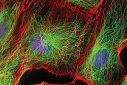
These human epithelial cells were labeled with fluorescent markers specific for microtubules (green), actin filaments (red) and DNA (blue). Microtubules and actin, components of the cytoskeleton, are responsible for cell structure, motility and division. Courtesy of Jennifer Waters Shuler, Nikon Imaging Center.
An example of science driving the technical advances is spinning-disc confocal microscopy. When it became available for biological microscopes, researchers let manufacturers know what they needed so as to use this new science consistently, and manufacturers responded with special optics, lighting, highly accurate focus capability, sensitive cameras and software.
The following articles describe how different types of microscopy are advancing, and what products are being developed to meet those demands. Prior Scientific, for example, brings automated shutters and focus mechanisms to the lab, allowing ongoing light microscopy studies to be performed as simply and accurately as possible. Also of note are recent developments in total internal reflection fluorescence microscopy, into which Olympus America provides some insight. In another feature, Leica engineers explore recent developments in fluorescence and forensic microscopy.
Rounding out this series of features is a look at adaptive optics, which was once primarily a tool for astronomers but is now making its way into microscopy, where researchers use it to counter aberrations in their systems.
Scientists at the University of Strathclyde’s Institute of Photonics in Glasgow, UK, have done extensive research into adaptive optics in microscopy, and those results are reported here, along with a glimpse of what might be available commercially in the near future.