Size and positioning demands push new stage designs.
David Denu, Ludl Electronic Products Ltd.
The axiom “Specialization is a necessary consequence of ubiquity” holds more truth today than ever for microscope positioning and motorized stages. Applications such as live-cell studies, image tiling, virtual microscopy and time-lapse studies are challenging microscope positioning technologies and driving novel, specialized capabilities.
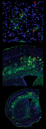
Images from a virtual slice montage represent more than 4000 fields of view of a mouse brain scanned using a 603 objective lens, three channels in confocal mode and Neurolucida DSU Virtual Slice software from MicroBrightField Inc. The high-power view shows the full resolution of the specimen. At medium power, the specimen appears as if it were viewed with a 103 objective. The low-power, macro view shows the specimen as if it were seen with a 0.53 objective lens. Courtesy of MicroBrightField Inc., Williston, Vt.
The challenges posed by these applications require an increasing degree of precision and accuracy. However, improving the performance of microscope stages is constrained by their small working envelopes and by size and weight limitations. Now, high-resolution, miniature versions of optical linear encoders help satisfy the demand for smaller microscope stages. This enabling technology has made the stage an essential part of the instrument, capable of extending its capabilities.
An example is a new stage system that directly embeds linear encoders in the stage rather than adding them as an option. By embedding the encoder, all of the performance gains are realized without additional cabling, brackets or fixtures that would increase the size of the stage or restrict the microscope function. In fact, the stage appears to be no different from nonencoder versions.
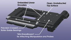
New applications challenge stage design. This flattop stage features high-accuracy linear encoder positioning with an unobstructed flat surface for freedom to mount apparatus for live-cell studies.
Some applications, such as time-lapse experiments, are very demanding with regard to stage stability and position reproducibility. With experiments that can run as long as minutes, hours or days, stage drift and other errors create uncertainty and adversely affect experimental results. A stage equipped with linear encoders offers much better stability, precision and overall performance. The high-resolution position feedback compensates for — and can often eliminate — accumulated errors. Typical bidirectional position repeatability is better than 0.25 μm. Nevertheless, linear encoders are not a panacea for poor design. Stage and component construction are still very important.
Live-cell applications for the study of mammalian specimens often involve environmental chambers, large specimens and sometimes an integrated manipulator, typically using an inverted microscope. The common inverted positioning stage, however, is not well suited for this application. A motorized inverted stage configuration, for example, has motors and drive housings placed so that specimen access and attachment options may be restricted. Furthermore, manipulators required for patch clamping and other tasks are impossible to mount.
A specialized solution is a flattop design, which provides a completely unobstructed platform to accommodate any size chamber or specimen holder easily and without interference. The stage also incorporates a unique “mini breadboard” area with a standard pattern of tapped holes for mounting manipulators and probes.
Stages for image tiling
In another extremely demanding application for microscope positioning stages — image tiling — a high-resolution image is created by linking smaller images, or tiles, into a mosaic. To help create the mosaic, the stage becomes, in effect, an extension of the microscope image. The performance of the stage is critical.
In tiling, stage precision determines how well adjacent images are registered. For example, at high magnifications and where the imaged field is ≤350 μm, stage accuracy must be as good as possible to ensure proper registration. Image overlap results in the duplication of features; conversely, gaps between images can be confusing in postprocessing. Software methods have been used to compensate for stage error by matching, warping, stretching and stitching the images tiles, resulting in images that appear to be continuous, free from error and suitable for presentation. However, uncertainty arises when quantitative analyses are made using the corrected data.
Similarly, in virtual microscope applications, an entire slide is scanned at high resolution. Very large high-resolution images can be scanned using software that enables networked virtual scanning of slides at different magnifications in a simulated microscope environment.
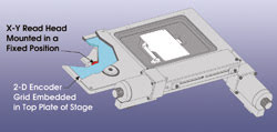
The requirements of image tiling applications demand high-precision positioning. This grid-encoded stage provides direct feedback reference to a precision X-Y grid embedded in the stage.
Ludl Electronic Products Ltd. designed a microscope stage specifically for use in image tiling applications. It requires no corrective software, is accurate to better than 2 μm over the full 100-mm travel range and has measured repeatability better than 0.2 μm. A two-dimensional grid encoder, which uses an orthogonal high-resolution X-Y line-space pattern on a glass plate, provides feedback on positioning.
The microscope stage is constructed so that the grid moves as the specimen moves. A single read head is mounted to a fixed position. The result is that, for any given position of the stage, the encoder will report both X- and Y-axis changes. This encoder system improves the performance of the stage to provide high accuracy, repeatability and, accordingly, research quality.
With new encoder technology and specific design improvements, stages can meet the demands of leading-edge microscopy. Nonetheless, we can expect new challenges as imaging technology and methods continue to develop. New applications that use cameras with higher sensitivity and data rates will further define performance in terms of speed, throughput and interconnectivity as well as accuracy and precision.
Meet the author
David Denu is vice president of marketing at Ludl Electronic Products Ltd. in Hawthorne, N.Y.; e-mail: [email protected].