
Standard PET/CT sometimes falsely detects cardiac problems
PET/CT scans are used to assess the severity of coronary artery disease and to follow its regression or progression. The images depict the amount and size of the abnormalities present and help physicians make appropriate treatment decisions. However, researchers recently discovered that standard PET/CT cardiac imaging often produces false-positive results.
As reported in the July issue of the Journal of Nuclear Medicine, Dr. K. Lance Gould from the University of Texas Medical School in Houston and his colleagues examined why and how often errors occur with standard cardiac PET/CT imaging. Two-hundred and fifty-nine patients being tested for potential coronary artery disease underwent cardiac PET/CT imaging with a 16-slice scanner from GE Healthcare. They were scanned at rest and after injection of dipyridamole, a drug that prevents blood clotting.
The first 145 patients then were imaged either during normal breathing or at the end of an expiratory breath-hold using helical CT attenuation correction, which is software intended to correct for the amount of attenuation. However, sometimes faulty defects appeared in the PET images because of the changes in thoracic-diaphragmatic structures during breathing that were not accounted for in the helical CT attenuation corrections. This happens because the helical CT scan lasts for only a few seconds and takes a snapshot of the structures at some moment in the respiratory cycle that may not match the average position of the thoracic structures of the many breathing cycles during the 6-min PET emission data acquisition.
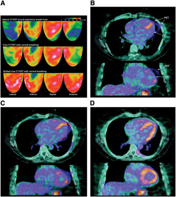
Researchers recently discovered that PET/CT cardiac imaging often produces false-positive results. However, PET/CT imaging used with cine CT attenuation correction provides more accurate results (see image A with helical CT-PET, cine CT-PET and the normalized images with shifted cine CT-PET). Also shown here are images (of the same patient as in A) of helical CT-PET with some marked misregistration (B), cine CT-PET with some misregistration (C) and shifted cine CT-PET with no misregistration (D).
The other 114 patients, in addition to the helical CT attenuation, underwent cine CT with attenuation corrections averaged during breathing by fusing the cine CT images together throughout the breathing cycle. The researchers compared the cine CT attenuation-corrected PET images with the standard helical CT-corrected PET images to understand and correct for the changes during breathing.
They found that misregistration of standard helical CT/PET images caused PET defects to show up in 40 percent of the patients. However, they also found that the PET images made with cine CT attenuation correction software normalized the images and gave accurate results.
The researchers believe that the results indicate that misregistration of PET/CT images with standard helical CT attenuation correction can cause false-positive results, but when used with cine CT attenuation correction can provide more accurate results.
Published: September 2007