“Microspectroscopy is basically the ability to obtain spectra of microscopic samples or sampling areas. It differs from other microscopy techniques in that it is primarily used to acquire spectra rather than images,” according to Paul Martin, president of Craic Technologies Inc. Based in San Dimas, Calif., the company specializes in microscopy and microspectroscopy in the UV-VIS-NIR regions.
Martin noted that both spectra and high-resolution digital images can be acquired with the company’s microspectrophotometers, also known as microspectrometers (including its QDI 2010 model), which are designed to measure the UV-VIS-NIR spectra of microscopic samples or microscopic areas of larger objects. Of the two types of instruments available, the fully integrated microspectrometer has been built for microspectroscopy, while the microscope spectrophotometer unit has been designed to attach to an open photoport of an optical microscope. Depending upon the configuration, microspectrophotometers can nondestructively measure the spectra of samples, even at the submicron level, by transmission, reflectance, fluorescence, and electron and polarization microspectroscopy, according to the company.
Applications for the UV-VIS-NIR regions are numerous, ranging from forensic sciences to geology to materials science, biology and pharmacology. “A lot of microspectroscopy is also done in the semiconductor and photovoltaic fields for everything from film thickness measurements to contaminant analysis,” Martin commented.
He noted that, although Craic Technologies specializes in integrating optical microscopy with optical spectroscopy, he has seen spectrometers of various types added to confocal, scanning electron and IR microscopes, and that Raman microspectrometers are also quite prevalent.
“Among the challenges faced in the microspectroscopy field are the problems that come with combining the operation of a microscope with a spectrophotometer, and the fact that you have a lot less light to work with at the microscopic level,” Martin said. “To acquire good-quality spectral data easily, it takes a well-thought-out design in terms of optics, hardware and software. Engineers are always trying to improve sensitivities as well as ease of use,” he added.
“When purchasing an instrument system, it is important to consider the entire integrated tool, and not just the components,” Martin explained. “As an example, the theoretical spectral range of the detector will be far larger than that of the instrument once you add a monochromator, optics, light sources and even the sample – yet the spectral range of only the detector is often quoted.”
Microspectroscopy provides the ability to transfer most spectroscopy methods to the microscope world, said Uwe Ortmann, head of sales and marketing at PicoQuant GmbH in Berlin. Spectroscopic techniques in common practice will be transferable to microscopic or even nanoscopic dimensions. The research will evolve to encompass the study of smaller and smaller samples, environments and particles. Confocal microscopy is commonly combined with time domain studies and intensity fluctuation (fluorescence correlation and cross-correlation spectroscopy), he said, adding that the company’s customers are also able to combine atomic force and confocal microscopy. Applications for microspectroscopy currently dominate in cell biology and protein research, mostly at the fundamental level.
A system for microspectroscopy, PicoQuant’s MicroTime 200 confocal time-resolved microscope is a high-end research tool for lifetime imaging, fluorescence correlation and single-molecule detection. It offers attomolar sensitivity down to the single-molecule level, Ortmann said, adding that the company is dedicated to making the instrument systems more user-friendly and lowering their costs.
Systems and advantages
In late November 2009, Craic Technologies announced the compatibility of Windows 7 computer programming with its Minerva microspectrometer control and spectral analysis software. The company says that scientists will notice a more fluid response with the program’s enhanced stability and advanced memory management. Windows 7 will further improve the usability of the software with features such as the quick resizing of windows, easier-to-see icons, speedy access to often-used documents and spectra, and a fast search engine to locate and quickly analyze data.
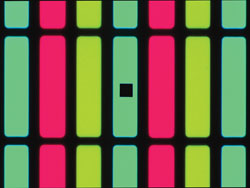
This is an image of how an organic LED chip appears under a microspectrometer when a spectral measurement is being made. The black square is the entrance aperture of the spectrometer.
In 2009, Craic Technologies noted that its QDI 2010 microspectrophotometer, combining both UV microscopy spectral imaging and the analytical capabilities of UV-VIS-NIR microspectroscopy, can be configured to help manufacturers locate and identify organic and inorganic contaminants in hard disk drive components such as read-write heads. Techniques commonly used for this purpose, such as inspection with optical microscopes, have not been able to adequately detect or analyze contaminants, according to the company.
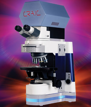
Craic Technologies’ QDI 2010 microspectro-photometer is a state-of-the-art instrument for UV-VIS-NIR microspectroscopy in the fields of forensics, materials science, biology, drug development and geology, according to the company. Photos courtesy of Craic Technologies Inc.
Craic Technologies also announced that its UV microspectrophotometers, such as the QDI 2010, can rapidly differentiate DNA from salt and even protein crystals by absorbance microspectroscopy – and that they can also qualify the crystal once it has been located and identified. Study of the structure of DNA is commonly done with x-ray crystallography, which requires the growth of DNA crystals that are stable and free from protein contaminants.
The company says that, using normal microscopy techniques, it is difficult even to identify DNA crystals, much less determine whether they are viable. DNA readily absorbs light at 260 nm, but a salt or protein will not absorb light at that wavelength. Due to the inherent flexibility of microspectrophotometers, besides imaging the crystals themselves, they can produce microspectra of crystals as small as a micron. The ability to use microspectra to qualify the crystal can save valuable time by enabling the selection of only viable crystals for the next step of a growth process.
Craic Technologies’ microspectroscopic technologies and techniques have applications in areas such as vitrinite coal analysis, measurement of surface plasmon resonance on the microscopic scale, and the rapid and accurate metrology of organic LED and liquid crystal displays for color, intensity and mura.
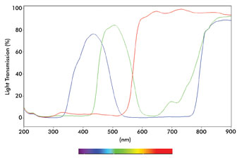
Shown is an overlay of three spectra from the organic LED chip (but with three different-colored pixels).
FTIR microspectroscopy applications
Biochemical changes associated with prostate cancer can be discriminated by Fourier transform infrared (FTIR) microspectroscopy to classify organ-confined and locally invasive prostate cancers, according to scientists at the Molecular Imaging Program at Stanford and the Stanford Infrared Optics and Photomedicine Center, both at Stanford University in California. The group compared the method with histological pathology for the evaluation of tissue for improved prostate cancer diagnosis and treatment. A report on the study appears in the December 2009 issue of Trends in Biotechnology.
Published by Thermo Fisher Scientific Inc. of Waltham, Mass., Application Note: 51517, titled FT-IR Microspectroscopy in Forensic and Crime Lab Analysis, covers the use of Thermo Scientific’s Nicolet iN10 infrared microscope – an optical microscope with integrated Fourier transform infrared instrumentation – in detecting counterfeit money, and in analyzing hair fiber, and tablet, paint and fingerprint residue.
Other methods and applications
In the area of Raman microspectroscopy, Craic Technologies’ CTR-1 MicroRaman spectrometer performs micro Raman spectroscopy rapidly by standard Raman scattering or by surface-enhanced Raman spectroscopy. Spectra of microscopic images are acquired while the user views them with a high-resolution digital imaging system. The device has applications in such areas as biological research and semiconductor metrology.
Published in the January 2010 issue of the Journal of Synchrotron Radiation, an article by F. Hahn and C.A. Melendres discusses their work using synchrotron IR reflectance microspectroscopy to study film formation and the breakdown of copper.
Developments and applications of soft x-ray spectromicroscopy with a focus on scanning transmission x-ray microscopy are discussed in an article written by Tae Hyun Yoon, a researcher at Hanyang University in Seoul, South Korea. Published in Applied Spectroscopy Reviews in March 2009, the article notes that, as a result of significant advances in x-ray optics and the greater availability of third-generation synchrotron sources, this spectromicroscopic technique has become an important analytical tool in several disciplines, including the environmental and materials sciences.