It is now more than 180 years since the first image was captured by Joseph Nicéphore Niépce from an upstairs window on his estate in France using pewter plates and a camera obscura – the forerunner of today’s cameras.
Images today are produced in myriad ways – using infrared, fluorescence, bioluminescence, x-ray machines, optical coherence tomography (OCT), lidar – for use in the medical, scientific and business industries; for space exploration; and for security purposes by the military and governments.
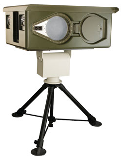
The T5000 is an outdoor people-screening system that can detect concealed threats at distances suitable for operational requirements. It can be used for checkpoint security, urban surveillance and counter-terrorism applications.
One of the most important breakthroughs in optical imaging in recent years is the development of CCD sensors, which laid the foundation for digital photography. Inventors of the CCD, Willard Sterling Boyle and George E. Smith, were duly recognized in 2009 with the Nobel Prize for physics.
Important advances also have occurred in the way we process and deliver images, such as compression technology used in digital TV, image processing associated with medical imaging and diagnosis, and the development of LCD displays for consumer electronics.
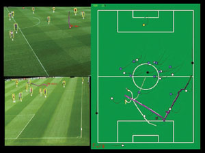
Image processing can help track players and a football during a match, to convey game information to a mobile phone. Courtesy of Digital Imaging Research Centre.
Medical imaging: Tools of the trade
Today, the most significant optical imaging tool used in medical imaging is still the standard optical microscope, which is used to analyze excised tissue. Tens of millions of biopsies are analyzed annually by trained histopathologists using these microscopes. However, it is not medical imaging in the same way as x-ray, MRI or CT, which can provide real-time images of a patient noninvasively.
Optical endoscopes are used widely for looking inside the body. But unlike x-ray and MRI, they do not provide a subsurface image. Another imaging method gaining momentum is confocal endoscopy via handheld probes or endoscopes. This technology provides high-resolution subsurface images but a very small field of view and very limited depth – up to approximately 250 µm.
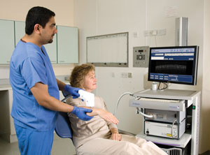
These images show Michelson Diagnostics’ VivoSight multibeam OCT scanner in action at University College Hospital in London. This product is not currently approved for clinical use in the USA. Images courtesy of Michelson Diagnostics.
OCT, undoubtedly a groundbreaker in terms of optical imaging, also is widely used in ophthalmology. In recent years, incremental improvements in the design of component light sources and probes have been opening the door to new applications; e.g., OCT imaging is expected to become widely used in diagnosing many types of cancer and in guiding treatment. It is making rapid progress in cardiology (scanning arteries for vulnerable plaques) and as a general-purpose imaging tool in dermatology.
“The combination of high-resolution, real-time two- and three-dimensional imaging capability, ease of use as well as nonionizing properties make it of great interest to clinicians,” said Jon Holmes, chief executive officer of Michelson Diagnostics Ltd. in Kent, UK. “In most cases, the depth of penetration is sufficient to penetrate mucosal layers to reveal details of organs and organelles that are too small to be seen with other imaging modalities and too deep for conventional microscopy.”
As volumes rise and component costs fall, Holmes believes that OCT also may make progress in price-sensitive applications such as dentistry – for early diagnosis of tooth decay and gum disease.
“Commercial growth will take off rapidly once OCT has ‘conquered’ a couple more clinical applications – as it has already done so in ophthalmology,” he said. “It will be seen that transferring the knowledge gained into new applications is just a matter of engineering and clinical testing.”
Terahertz imaging: New applications
Astronomical imaging has always been a major driver of innovations in optics. New imaging technology came to life when the European Space Agency’s StarTiger team captured the world’s first terahertz-frequency picture of a human hand in September 2002.
Terahertz waves, defined as frequencies from 100 GHz to 100 THz, easily can pass through some solid materials, such as walls and clothes, penetrating materials that are usually opaque to both visible and infrared radiation. This makes terahertz cameras ideal for security applications, where they potentially could be used to screen airport passengers for hidden objects.
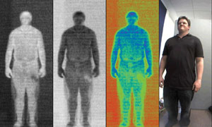
Screening people for concealed objects is just one of the promising applications of terahertz radiation. Courtesy of ThruVision Systems Ltd.
For the screening of dangers of a different kind, terahertz also is showing great promise in medical imaging. Light at this part of the spectrum is absorbed strongly by large biological molecules and by water and is subject to far less scatter than visible or infrared wavelengths, resulting in sharper images. Furthermore, unlike x-rays, terahertz radiation is nonionizing, so there are no safety issues to consider.
Technology operating in the terahertz regime is still in its infancy compared with the rest of the electromagnetic spectrum. This is largely a result of the “terahertz gap” between solid-state electronic devices and photonic devices. Despite this, a growing number of firms are catching on to the untapped potential of terahertz technology.
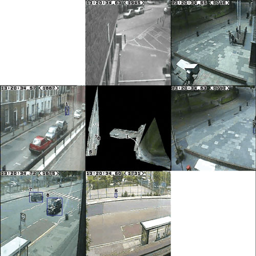
Tracking people and vehicles across various cameras can be useful in interpreting movement for public closed-circuit television systems or fortraffic monitoring. Courtesy of Digital Imaging Research Centre.
“Our current products all image passively at 1250 microns,” said Chris Mann, chief scientific adviser at ThruVision Systems Ltd. in Abingdon, UK. “With respect to passive imagery, this part of the electromagnetic spectrum is completely unexplored, and as the imaging equipment becomes more widely available, we are convinced many new applications will emerge.”
Companies such as ThruVision are helping terahertz advance into other fields. One of the first to emerge is in production and quality control. Infrared cameras are used routinely in this sector, but for some materials, their limited penetration means they cannot meet industry needs. In contrast, terahertz cameras can see through great thicknesses – 10 to 1000 nm – of many common low-density manufacturing materials to spot unwanted defects, such as voids or inclusions, in real time.
Digital imaging, processing
Enhancing and extracting features of interest are all part and parcel of image processing. Increasingly powerful computers have enabled image processing to be performed in real time, opening up many ultramodern applications.
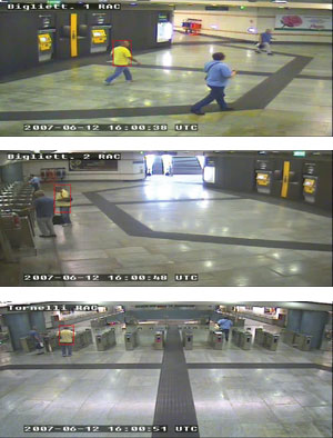
These images track the same person across many cameras for public closed-circuit television systems. Courtesy of Digital Imaging Research Centre.
“Much of the human brain is used to process visual information so that we can make decisions, explain, archive and so on,” said Dr. Sergio Velastin, director of the Digital Imaging Research Centre at Kingston University in the UK. “To be able to do the same automatically using computers is an important goal for a wide range of practical applications, including surveillance, driverless trains, unmanned space exploration, automatic tagging of contents and advanced robotics, to name a few.”
Digital imaging is being adopted increasingly in manufacturing industries, where it is used in machine vision systems for sorting, quality control and assembly. But one of the most intriguing applications lies in motion capture.
Motion-capture systems can be used to track and interpret movement – be it human behavior for public closed-circuit television systems or vehicle motion for traffic monitoring. “Understanding human/vehicular behavior helps to prevent incidents,” Velastin said. “And detecting anomalies in normal behavior means better controls can be put in place.”
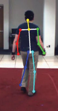
Extracting a person’s “pose” using a single camera can be used to assess improvement in a patient’s body movement after an operation or physiotherapy. Courtesy of Digital Imaging Research Centre.
Motion capture also is making waves in medicine, where it can be used to assess improvement in body movement after physiotherapy or an operation, and it is finding application in the gaming industry to create virtual worlds.
The possibilities may seem endless, but according to Velastin, investors must be more adventurous to fund companies that will encourage end users and suppliers to embrace the potential of imaging. An idea of what we could expect in the next five to 10 years includes assisted living for the elderly or infirm, driver-assisted systems in the automotive industry, and intelligent home/office environments that respond according to human presence and motion.