
MEMS-based scanning device facilitates microendoscopy
Gwynne D. Koch
Line-scanning rates of up to 1 kHz are important for imaging fast
biological processes such as blood flow and neuronal activity. However, conventional
scanning mechanisms that offer fast acquisition rates, including galvanometer, spinning
polygon and acousto-optic scanners, cannot readily be miniaturized for incorporation
into microendoscopes for minimally invasive imaging procedures.
Most miniaturized scanners explored for confocal
and two-photon fluorescence imaging have been cantilever mechanisms, such as a vibrating
optical fiber mounted on a piezoelectric actuator. Although inexpensive to fabricate,
these mechanisms offer limited scanning rates, are not easily mass-produced and
cannot be reduced to millimeter sizes.
Now, a team of optical scientists and
engineers led by Mark J. Schnitzer, Olav Solgaard and Wibool Piyawattanametha at
Stanford University in California has developed a device that is based on microelectromechanical
systems (MEMS) scanners and that achieves adjustable, fast-axis acquisition rates
of up to 3.52 kHz.
Using reactive ion etching methods,
the researchers fabricated 750 x 750-μm single-crystalline silicon scanning
mirrors on a double silicon-on-insulator wafer. Six banks of electrostatic vertical
comb actuators and a gimbal design enabled rotation of the scanning mirror in two
dimensions with minimal crosstalk. The mirror, movable comb teeth and
inner torsional springs were fabricated in the upper device layer, and the frame,
fixed comb teeth and outer torsional springs were fabricated within both the upper
and lower layers, each measuring 30 μm thick.
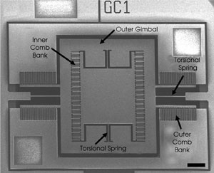
An electron micrograph shows the key components of a two-dimensional
MEMS scanner developed for two-photon microscopy and microendoscopy applications.
Images reprinted with permission of Optics Letters.
The layer thickness is important because
it affects the performance of the MEMS scanner; thicker mirrors can reduce flexure
of the mirror when scanning it at high speeds, and the pronounced thickness of the
comb teeth raises the electrostatic torque that can be applied to the mirror, increasing
its angular range to up to 16°.
To test the feasibility of optical
imaging based on MEMS, the scientists constructed a two-photon microscope that employed
their scanner. A Spectra-Physics Ti:sapphire laser provided an 850-nm excitation
beam with a pulse width of 100 to 150 fs and a repetition rate of 80 MHz. The beam
passed through two lenses that decreased its diameter before reflecting off the
scanner, then expanded and passed through a dichroic mirror until it filled the
back aperture of the microscope objective. Fluorescence was detected with a Hamamatsu
photomultiplier tube. The instrumentation can be additionally equipped with a compound
gradient refractive index (GRIN) microendoscope probe, placed after the objective
lens, for microendoscopy applications.
Using two-photon microscopy and microendoscopy,
the team captured images of pollen grains with micron-scale detail. Acquiring
data on both the forward and return scanning paths enabled acquisition rates of
up to 3.52 kHz.
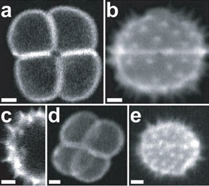
Two-photon fluorescence images of pollen grains were captured using instrumentation incorporating
the MEMS scanning mirror. Images were acquired using a microscope objective (a-c)
or a GRIN microendoscope probe (d,e).
Given the measured ranges of the two
axes and 850-nm excitation, the number (N) of distinguishable focal spots
for any imaging system based on the researchers’ MEMS scanner is ~250 x 90.
The largest field of view that can be obtained without sacrificing imaging resolution
(R) is about N x R. Given the highest lateral resolution demonstrated
for GRIN endoscope probes, ~1 μm, the MEMS scanner offers a maximum field of
view of 250 x 90 μm. For typical microscopy applications in which R = ~0.5 μm, fields of view of ~125 x 45 μm are obtainable.
The value of N for the MEMS
scanning mirror is smaller than that of galvanometer mirrors and other scanning
mechanisms. Modest gains in N might be achieved by increasing the maximum
scanning angle or the diameter of the mirror, but the latter option would decrease
the mirror’s scanning speed. The researchers are working on incorporating
the MEMS scanner into a miniaturized fiber optic instrument to be used for portable
two-photon microendoscopy.
Optics Letters, July 1, 2006, pp.
Published: September 2006