To see something small, you need a magnifier
— and that is what a group of researchers at the Georgia Institute of Technology
in Atlanta turned to in developing a label-free biosensor. For their work, they
used hydrogel microlenses to monitor specific protein-binding events.
A hydrogel is a network of water-soluble polymer
chains. When coated with an antigen and placed in the presence of the appropriate
antibody, the hydrogel changes size because of the antigen-antibody binding. In
theory, a suitably coated hydrogel particle could detect the presence of a specific
bioagent. In practice, however, it is difficult to measure the small amount of swelling
in such tiny particles.
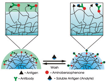
In this label-free biosensing scheme, antigen-coated hydrogel microlenses
are assembled on a glass substrate and functionalized by photochemically adding
antibodies to their outer layer. The antigen-antibody binding causes the lenses
to swell, detectably changing their focal length (right). Photoactivation of aminobenzophenone
using UV radiation holds the anti-biotin onto the hydrogel after binding. The swelling
is reversible by washing the hydrogel. Reprinted with permission from Angewandte
Chemie.
Several years ago, the researchers
realized that hydrogel particles adsorbed to a surface could be induced to bead
up and form hemispherical lenses. When the particles swelled, the focal length of
the lenses changed, providing a very sensitive measure of a change in the surface.
In a demonstration of this, they prepared
hydrogel microparticles of approximately 2 μm in diameter on a glass slide,
coating the particles with the antigen biotin. Using a photochemical process, they
coupled biotin antibodies to the outer layer of the coated hydrogel particles. To
measure an individual lens, they projected a pattern through it, defining it as
being “on” if the pattern was faithfully reproduced. They also used
differential interference contrast microscopy, defining the lens as being on if
a dark ring appeared around the particle’s periphery.
L. Andrew Lyon, an associate professor
of chemistry and biochemistry at the institute, said that the technique might have
room for improvement, and that this is the focus of current research. “Right
now, we are using simple optical microscopy to monitor focal lengths, and we frankly
do not know if that is the most sensitive method or not,” he said.
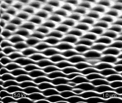
Scanning electron microscopy depicts an array
of the hydrogel microlenses. Courtesy of L. Andrew Lyon and Georgia Institute of
Technology.
For their demonstration, the researchers
exposed their microlenses to a solution containing the antigen biocytin and measured
the effect. They then washed the microlenses with the appropriate solution to free
the antigens — restoring the particles to their original state — and
measured them again. They repeated this cycle several more times.
In a series of tests, they showed that
the sensor could reliably detect the antibody at concentrations of about 10 nM.
This was a binary reading, with the sensors indicating the presence of the analyte
but not its concentration. However, they could and did change the point at which
this transition occurred by adjusting the antibody concentration in the periphery
of the hydrogel.
Unlike some more traditional bioassays,
their technique detects the analytes in seconds, not hours. Because the sensing
elements measure a couple of microns across, millions can be packed into an inch-square
plate. That density could be useful, Lyon noted.
“One could envision a microlens
array where groups of elements would be responsive to a specific analyte and different
members of that group would respond to different concentrations,” he said.
Besides improving the measurement method,
the researchers seek to demonstrate more sensitive sensing elements, to fabricate
multianalyte arrays and to integrate the technique into microfluidics so that sampling
can be well controlled. “We are also investigating different synthetic methods
for creating microlenses that display more dramatic changes in focal length,”
Lyon said.
He added that fabrication of bioresponsive
materials is important in areas beyond bioanalysis and that these areas also are
being investigated.
Angewandte Chemie Int Ed, Feb. 20, 2006, pp. 1446-1449.