Photographers know that good photos of children and pets can involve certain forms of coaxing: For children, you tell them to say “Cheese!” because it will look like they’re smiling; for pets, you bribe them with tasty treats to pose for a sweet second or two.
Dr. Igor Siwanowicz’s subject required a very special kind of treat: single-celled organisms. But the bribe paid off, in the form of first prize – $5000 worth of Olympus equipment and serious bragging rights – in the 10th-anniversary Olympus BioScapes Digital Imaging Competition. The winners were announced at a reception during the recent American Society for Cell Biology annual meeting in New Orleans.
Siwanowicz, a researcher at the Howard Hughes Medical Institute’s Janelia Farm Research Campus in Ashburn, Va., used confocal microscopy to capture the floating humped bladderwort plant as it consumed a “mouthful” of microinvertebrates. The image beat out more than 2100 entries.
The subject, an aquatic carnivorous plant called Utricularia gibba, sucks the organisms into its trap and digests them a millisecond after they touch its trigger hairs (seen in the center of the image, inside the dome-shaped entrance). The traps also provide a microhabitat for single-cell green algae, predominantly desmids, which are visible in the photo.
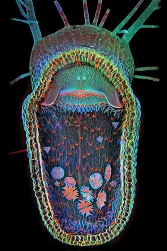
This image of an aquatic carnivorous plant with tiny organisms inside its maw won Dr. Igor Siwanowicz first prize in the 10th-anniversary Olympus BioScapes Digital Imaging Competition. Courtesy of Siwanowicz/Olympus Bioscapes.
The annual competition honors images and movies of life sciences subjects – human, plant and animal – as captured by light microscopy. Judging is based on the science depicted in the images as well as their beauty or impact, and the technical expertise involved in capturing them. Besides the top 10 award winners, 69 honorable mentions were named this year, including 55 still images and 14 movies. A wide variety of microscopy techniques contributed to the top 10 in 2013: dark field, structured illumination, polarized light, confocal, fluorescence, stereomicroscopy and more.
This year’s winners exemplify advances in neuroscience and cell biology research, or offer glimpses of microscale life captured by creative hobbyists, students and other scientists. Dorit Hockman of the University of Oxford in England nabbed second prize for an image of a black mastiff bat embryo at the “peekaboo” stage, when its wings have grown to cover its eyes. Siwanowicz also won third prize for an image of desmids and earned an honorable mention for his image of rotifers surrounding a green alga.
“For 10 years, Olympus has sponsored this competition to bring to light the power, beauty and importance of science and the work that scientists do,” said Brad Burklow, executive director of international business for the Scientific Equipment Group of Olympus America Inc. “BioScapes movies and still images combine art and science to remind us of the fascination and wonder of the natural world and highlight vital work going on in laboratories. They serve as an inspiration to young people seeking careers in science, technology, engineering and mathematics.”
Next year’s competition is already open for entries; it closes Sept. 30, 2014.