
Interferometric Technique Targets Protein Shadows
A new optical method for targeting proteins could mean earlier diagnosis and more effective treatment for cancer.
The technique makes individual proteins — including those characteristic of certain types of cancer — visible and was developed by a team from the Max Planck Institute for the Science of Light. Using interferometric detection of scattering (iSCAT), the researchers directly detected the scattered light of individual proteins in cancer cells via their shadows.
In the study, the researchers shined laser light onto a microscope slide on which the relevant proteins were captured with appropriate biochemical lures. The proteins scattered the laser light, thus casting a shadow.
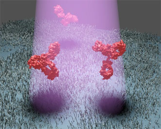
Researchers can identify individual proteins by the shadow produced when they scatter light. Courtesy of the Max Planck Institute for the Science of Light.
The new technique targets and produces a clear image of the proteins without the need for the traditional luminous markers.
“Until now it was thought that if you want to detect scattered light from nanoparticles, you have to eliminate all background light,” said professor Dr. Vahid Sandoghdar.
“However, in recent years we’ve realized that it is more advantageous to illuminate the sample strongly and visualize the feeble signal of a tiny nanoparticle as a shadow against the intense background light.”
The researchers allowed the background light to interfere with the weak, scattered light so that the desired signal would be amplified.
However, they were initially unable to detect the shadows of a single protein in the interference image because the pattern was distorted by background noise. To eliminate the noise, the researchers took images of the microscope’s sample holder before and after it contained the protein. The target proteins then stood out clearly from the background as dark spots.
“Since most of the optical noise generated by nanoscopic irregularities of the sample do not change, we can subtract one image from the other and thus eliminate the noise,” said Dr. Marek Piliarik.
Via iSCAT, the researchers may also detect individual proteins from mixtures that contain concentrations of other proteins up to 2000 times greater.
“The specificity of the detection method is not limited by the optics but by the selectivity of the substances used to bind the target proteins to the microscope slide,” Piliarik said.
The researchers found that the amount of contrast between the protein and background depends on the size of the target particle. Such contrast could reveal information about the shape of the protein.
The technique could also allow the researchers to study how different proteins interact with each other, which is crucial for understanding diseases, as well as a broad range of biological processes, according to the researchers.
The work was published in Nature Photonics (doi: 10.1038/ncomms5495).
For more information, visit www.mpl.mpg.de.
Published: September 2014