Leading analytical and clinical diagnostics instrumentation OEMs rely on integrated light emission and detection solutions to unlock the mysteries around disease and treatment. Photonic solutions encompassing optical, illumination, sensing and optomechanical technologies provide these OEMs with not only convenience and simplicity, but also an accelerated path to market for the development of highly complex life science and analytical instruments. Even standard, off-the-shelf components, when coupled with a modular and expandable architecture design, can be used to create a fully functional flow cytometer that achieves levels of throughput and cell analysis sensitivity comparable to commercial products.
RICHARD SIMONS, EXCELITAS TECHNOLOGIES CORP.
Life science and analytical instruments based on optical techniques have moved from university and corporate research laboratories, through central diagnostic and pathology laboratories, to local hospital laboratories. They are now becoming more and more commonplace as diagnostic tools at or close to the point-of-care delivery. Enabling this development is the evolution of optical components and subsystems used within these instruments for the emission, manipulation and detection of light. The result has been increased capability and usability, along with decreased size and cost. For any optical instrument, the beam path can be summarized as follows in Figure 1.
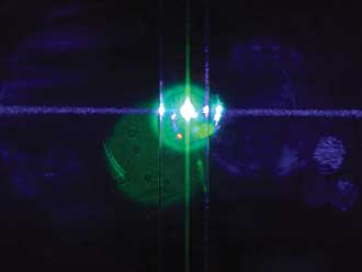
Laser-induced fluorescence in the heart of the flow cytometer. The laser beam is the violet beam, entering from left, inducing fluorescence in the target flow in the center of the flow channel, entering from top.
A good example of an analytical instrument is the flow cytometer, which will be used in this article to highlight the development of the optical heart of the analytical instrument system. Flow cytometers are instruments that measure features of cells in a liquid suspension, characterizing and analyzing cell populations using light scatter and fluorescence parameters. These life science instruments began as research laboratory instruments, and they still play an important role in research into immunology and cell biology. However, they are now also used in the clinical laboratory for diagnosis and monitoring of diseases such as HIV/AIDS and blood cancers. The next phase would be for them to become diagnostic tools used in the clinic or doctor’s office lab, right at the point-of-care.
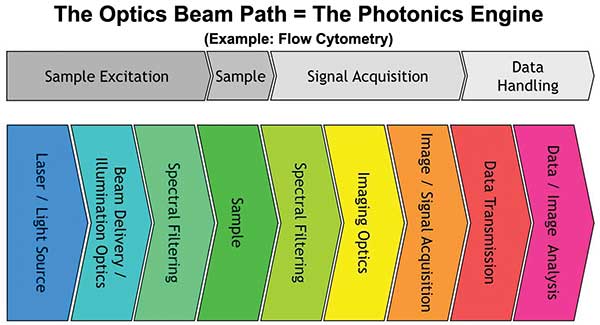
Figure 1. Block diagram of the optical beam path in a typical analytical instrument. Image courtesy of Excelitas Technologies Corp.
Movement toward smaller laser sources
Traditionally, the instrument manufacturer would have had to begin by mastering the photochemical reactions key to the instrument function. The manufacturer would also need to design the complete optical system within the instrument and then manage the sourcing of the necessary components from a large number of other manufacturers. The lasers originally used in the instrument would have been large, powerful argon or krypton ion lasers, needing water cooling and three-phase high-voltage electrical supplies. The beam would have been delivered through a free-space optical system of lenses, filters and microscope objectives to shape the beam and deliver it to the flow cell. Collecting the fluorescent or scattered signal would also have been done using free-space components, including lenses and mirrors, bandpass filters, and complex optomechanical mounts for alignment, allowing the signal to be delivered to the detectors. These detectors would typically have been a photomultiplier tube, a fragile and complex structure of electrodes sealed in glass vacuum tube, requiring over 1000 V to convert the photons entering the detector into a useful electronic output.
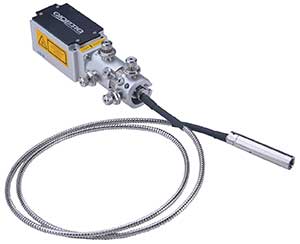
Qioptiq iRIS fiber-coupled laser module from Excelitas Technologies Corp. Photo courtesy of Excelitas Technologies Corp.
Today’s instrument designer can now use significantly smaller laser sources, using diode lasers and diode-pumped, solid-state lasers. These lasers can generate the wavelengths used to excite the fluorescence and provide the illumination for the scattering measurements. Usually, each excitation laser source is dedicated to one probe on the sample per run. Running parallel processes by using multiple illumination wavelengths fired in series on the sample can speed up the analysis.
Challenges in flow cytometry
In flow cytometry the primary challenge is to focus the light onto a moving sample in the flow stream, which is usually less than 100 microns wide. To gather meaningful data from a moving target, both the detector and illumination source need to be as stable and stationary as possible; otherwise movement from either one will cause image jitter and reduce resolution. The second physical challenge is positioning the beams from multiple light sources in sufficient proximity to generate parallel illumination spots focused in the flow cell, while sufficiently separating these spots to prevent cross-talk in the detection channels, which may collect emitted and fluorescent light simultaneously.
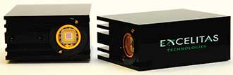
LynX silicon photomultiplier modules, as used in the flow cytometer demo system. Photo courtesy of Excelitas Technologies Corp.
Flow cytometers have traditionally used an optical system of lenses, filters and microscope objectives to shape the beam and deliver it to the flow cell. The principles are well-understood, although this can often result in a long optical beam train, making it more susceptible to the effects of laser jitter. An open beam path with optics mounted at various locations is also more susceptible to thermal temperature differences that will cause beam movement on the sample. Careful alignment of the optics is necessary as small movements can cause large changes in the beam pointing on the sample. As a result, instruments that use free-space optics can be susceptible to physical knocks and changes in environmental conditions, including heat generated by the laser itself.
The single-mode optical fiber solution
One way to guarantee a stable high-quality beam delivered to the sample without the use of complex optical components is to use single-mode optical fiber. This offers several advantages in functionality, from increased laser stability and image resolution to reduced instrument size and greater ruggedness.
Photo courtesy of Excelitas Technologies Corp.
Coupled directly from the laser to the fiber, the beam is much less sensitive to movement and thermal temperature changes, which reduces the need for instrument alignment service visits and creates a more robust instrument design. The fiber also acts as a spatial filter and eliminates any beam astigmatism to create a near-perfect Gaussian profile. The resulting beam is much more stable over time than those in free-space systems, and it is without accumulated errors from multiple optical interfaces. That helps ensure a more reliable and more stable measurement. Additionally, the use of fiber allows the laser source, which generates heat, to be located away from the flow cell. This means more flexibility for instrument layout, including the option of mounting the laser externally to the instrument head. It also simplifies servicing. That is because the optical alignment of the instrument is fixed and is independent of the laser — so there is no need to perform a lengthy realignment process when the laser is serviced.
Collecting the fluorescent and scattering signals from the sample requires the light to be gathered and collimated prior to using filters to select the wavelength area of interest. Again, this can be done completely in free space, or with the use of fiber, to simplify the optical set-up and to provide a stable delivery route to the detectors.
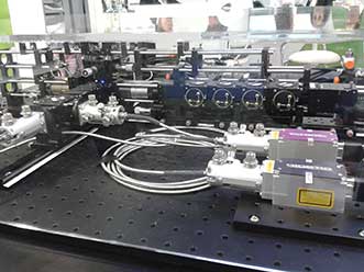
Photo courtesy of Excelitas Technologies Corp.
For detection of these signals, today’s designers can still use photomultiplier tubes (PMTs) for the ultimate in sensitivity. However, the advent of large area avalanche photodiodes (APDs) and silicon photomultipliers (SiPMs), allows a single detector type to support a wider range of scattered and fluorescent wavelengths. The result is a greater number of dyes can be used. An SiPM is a monolithic array of silicon micro-APDs offering high photon detection efficiencies from the near UV through to red and NIR wavelengths, coupled with relatively low dark counts. With low operating voltages compared to PMTs, as well as greater ruggedness and lower cost due to the solid-state nature of a silicon detector, many flow cytometer vendors are switching away from PMTs as their detection solution.
Simplifying the photonic engine design process
Many new and innovative analytical and diagnostic companies are using optical technologies such as flow cytometry to provide solutions to their customers. The goal of modern optical component suppliers is to make the manufacturer’s life easier by shouldering the burden of the complete photonic engine design, allowing the manufacturer to focus on its core competencies. To that end, demonstrating the ease and expediency of developing life science and analytical instruments from the ground up, Excelitas Technologies and its subsidiary Qioptiq decided to build a fully functional modular flow cytometry prototype. This prototype uses off-the-shelf products from Excelitas’ various divisions.
The design process began with lasers as excitation sources, incorporating two compact solid-state Qioptiq iFLEX-iRIS lasers, with one being blue and the other being violet. The violet laser took advantage of novel violet-excitable fluorescent dyes recently introduced for flow cytometry. The units were coupled with fiber optic delivery systems, allowing them to be positioned some distance from the sample for accessibility and an efficient design. They also simplified the alignment process and made for plug-and-play laser head replacements.
To capture and collimate the scattered and fluorescent light, two Optem High Resolution 20× microscope objectives were used. One was used for forward-scattered light (indicative of cell size) and the other was used for side-scattered light and fluorescence. Sets of longpass and bandpass filters ensured transmission of the appropriate light wavelengths to the detectors. Six high-performance SiPM modules were also used for photon detection, with one being used for the forward-scattered light, and a second for the side-scattered light. The other four detected the spectral emission wavelengths for fluorescence intensity, based on the lasers selected. The modules were SiPMs integrated with a power supply and amplifier, and they were simpler to integrate than conventional photomultiplier tubes or silicon components.
LINOS Microbench parts were used for organizing the optical bench. Anodized aluminum cubes with stainless steel posts, along with mounting plates, adaptors and connectors, were used with a compatible stainless steel rail system to secure small optical components and create the system layout. Microbench parts assemble and disassemble easily, yet they can be stably aligned, making impromptu adjustments to the layout practical. Cubes could be placed next to one another or stacked, simplifying the layout of components on all three axes. The system was also used to mount the fluidics and flow cell needed for a fully functional flow cytometer.
The system alignment was fine-tuned after assembly using fluorescence dyes and calibration particles. To show that the instrument met specifications, standard fluorescence reference particles were run on the completed system. This successful project clearly demonstrated the power of using standard, off-the-shelf components and of a design approach based on a modular and expandable architecture. This fully functional flow cytometer, designed and built from scratch in less than two months, achieved levels of throughput and cell analysis sensitivity comparable to commercial products.
From modularity and flexibility follows customization for reduced size and cost. That is what manufacturers must strive to achieve when striving to provide instruments — and not only cytometers — to users worldwide.
Meet the author
Richard Simons is the senior applications specialist with Excelitas Technologies Corp. in Montreal; e-mail: [email protected]