Using two-photon fluorescence microscopy, researchers have gained new insight into how the spinal cord mediates commands from the brain to get the body moving.
The findings could help identify ways to repair damaged neural connections in patients with spinal cord injuries or neurodegenerative diseases like amyotrophic lateral sclerosis (ALS), also known as Lou Gehrig's disease.
Researchers at the Salk Institute for Biological Studies used a fluorescent tag that could be added to multiple neurons and lights up whenever any of the cells are activated. Observing a mouse spinal cord through a microscope, the researchers could watch in real time which cells were activated when chemicals that turn on walking circuits were added.
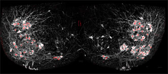
Scientists selectively added fluorescent proteins to the nuclei of motor neurons (red) to show how cells in the spinal cord synchronize many neurons at once to allow complex movements. Courtesy of Christopher Hinckley/Pfaff Lab, Salk Institute.
"You don't need to do any kind of post-image processing to interpret this," said Samuel Pfaff, a professor in Salk's Gene Expression Laboratory. "These are just raw signals you can see through the eyepiece of a microscope. It's really a jaw-dropping kind of visualization for a neuroscientist."
Pfaff's group used the new method to answer a long-standing question about how a collection of cells in the spinal cord, called the locomotor central pattern generator (CPG), connects to the right motor neurons to allow movements like walking.
The CPG is where relatively simple signals from the brain are translated into more complex instructions for motor neurons to control muscles. But until now, researchers didn't know exactly how CPG cells forged connections with motor neurons.
By tweaking the locations and identities of motor neurons and then watching the resulting patterns of activation, researchers found that CPG cells didn't rely solely on motor neurons' locations to connect to them. Instead, the genetic identity of each subtype of cells — what makes those that control the quadriceps muscle different from those that control the calf muscle, for instance — is also important.
That's a key finding for research on how to treat spinal cord injuries and ALS, Pfaff said.
Current efforts involve stem cells that are transformed into motor neurons and then implanted into the spinal cord to regenerate damaged connections. The Salk team's results, however, suggest that general motor neurons might not do the trick; specific subtypes of motor neurons may be necessary.
Funding for the work came from the National Institutes of Health, Howard Hughes Medical Institute, Christopher and Dana Reeve Foundation, Sol Goldman Trust and Marshall Heritage Foundation.
The research was published in Neuron (doi: 10.1016/j.neuron.2015.08.005).