Technology that combines the best features of two microscopy methods yields images 100 percent sharper than those acquired through conventional light-sheet-based microscopy (LSM).
LSM, also known as single-plane illumination microscopy (SPIM), uses a laser beam, narrowed to just a few microns across, to illuminate a biological sample from the side — instead of from above or below — with a thin sheet of light. A lens is then used to focus the fluorescence radiated from the sample upward to be captured by a digital camera.
A drawback of the method is that it enables only a portion of the sample to be imaged at a time. Rotating the sample, as well as raising and lowering the illumination plane, produces a series of two-dimensional sectional views, or “slices,” that can be pieced together to yield a 3-D map of a whole organism or any of its organs or systems.
The new method developed by a team in Italy allows high-speed, single-plane images of multiple sections of a sample to be taken while also eliminating the scattered background light that causes blurriness.
The new integrated LSM/confocal microscopy technique, called confocal light-sheet microscopy (CLSM), uses a filter to remove photons that stray from the thin sheet’s single plane.
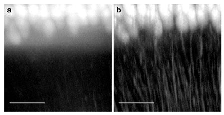
Purkinje cells from a mouse cerebellum imaged (a) with light-sheet microscopy and (b) with the significantly higher contrast provided by confocal light-sheet microscopy. The scale bar at the bottom is 100 µm across. (Image: Optics Express/European Laboratory for Non-Linear Spectroscopy, University of Florence, Italy)
Francesco Pavone, leader of a collaborative team comprising six Italian research agencies and an author of the paper describing the advance, said the combined systems “filtered the scattered photons that were emitted and recovered the normally lost image contrast in real time without the need for multiple acquisitions or any postprocessing of the acquired data.”
Researchers have tried to map the brain’s billionfold neural network with conventional LSM, but while the technique yields high-resolution views of tissue excised from mouse brains and those fixed in position, it cannot obtain whole-brain images. Whole-brain samples scatter the emitted light and create background fluorescence that reduces contrast and blurs the perceived image, an aberration that makes it difficult to resolve and reconstruct the entire neuronal network with high contrast.
The researchers say the new technique proved superior over conventional LSM microscopy and a variation of LSM that requires redundant slices and postprocessing to remove scattered light when used to view three samples of the mouse brain: the hippocampus, the cerebellum and the whole brain. They also used CLSM to map the micron-scale neuroanatomy of mouse Purkinje cells — large neurons found in the cerebellum — and to trace an entire brain’s neuronal projections.
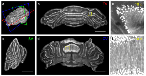
Micron-scale neuroanatomy of a whole thy1-GFP-M brain. (a) Isosurface perspective (P) and transverse (T), coronal (C) and sagittal (S) contours of an entire PND-15 mouse brain. The volumes highlighted by the blue and red boxes are magnified in panel (b) and (c), respectively. (b) Volume rendering of a portion of hippocampus. (c) Volume rendering of a portion of superior colliculus. (d) Soma segmentation and process tracing of selected fluorescent neurons present in the red box (c). For clarity, each neuron was drawn with a different color. Scale bars, 200 µm. (Image: Optics Express)
“We hope that the whole-brain tomographies we can obtain will one day provide insights into the mechanisms of these and other brain disorders.”
The study appeared in The Optical Society’s open-access journal Optics Express.
In other microscopy news, researchers at the University of Leicester have built a system that is 100 times faster. (See: Microscope Addresses the Need for Speed)
For more information, visit: www.lens.unifi.it