Photonics HandbookBioScan
Photoacoustic Microscopy Method Delivers Higher Sensitivity
A multispectral, super-low-dose photoacoustic microscopy (SLD-PAM) system developed by City University of Hong Kong (CUHK) achieves significantly higher sensitivity than traditional optical resolution photoacoustic imaging.
By providing an exceptionally high level of sensitivity, SLD-PAM could help broaden the use of photoacoustic microscopy in biomedical applications. In the future, it could translate to clinical settings; for example, it could be used for ophthalmic exams where a low-power laser is preferred for the patient’s safety and comfort. Long-term monitoring of pharmacokinetics or blood flow also requires low-dose imaging to alleviate perturbation to tissue function.
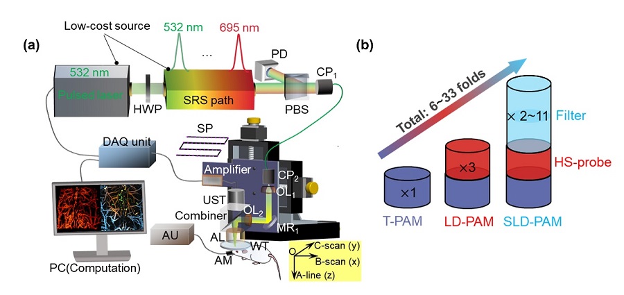
(a) SLD-PAM system and (b) sensitivity improvement from the probe and filter. Courtesy of Zhang, Y. et al., https://onlinelibrary.wiley.com/doi/10.1002/advs.202302486.
“High sensitivity is important for high-quality imaging,” said CUHK professor Lidai Wang. “It helps detect chromophores — molecules that confer color on materials by absorbing particular wavelengths of visible light — that do not strongly absorb light.”
Additionally, since a low-power light source is sufficient to operate SLD-PAM, it reduces the chances of photobleaching, phototoxicity, or perturbation to delicate tissues.
To build SLD-PAM, the researchers optimized the photoacoustic probe design of a technique they developed previously and implemented a spectral-spatial filter.
The researchers customized a high-numerical-aperture acoustic lens to optimize the optical and acoustic beam combiner and improve the alignment between the optical and acoustic foci. They combined these improvements with an innovative, 4D, spectral-spatial filter algorithm based on discrete wavelet transform. The algorithm enabled SLD-PAM to filter coherent photoacoustic signals in all 3D space and all-optical wavelengths, greatly enhancing the microscope’s ability to recover weak signals.
These improvements increased the sensitivity of the microscope by a factor of between 6 and 33, compared to the researchers’ previous technique.
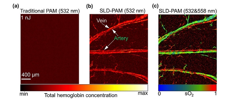
Comparison of the in vivo results of (a) traditional-PAM, (b) SLD-PAM at super-low pulse energy with a green-light source, and (c) oxygen saturation image acquired by SLD-PAM via dual-wavelength spectrum unmixing. Courtesy of Zhang, Y. et al., https://onlinelibrary.wiley.com/doi/10.1002/advs.202302486.
SLD-PAM uses a low-cost, multiwavelength, pulsed laser that provides 11 wavelengths ranging from green to red light. The laser operates at a repetition frequency up to megahertz, and the spectral switching time is in submicroseconds.
The researchers tested SLD-PAM through in vivo animal imaging at super-low pulse energy with green-light and red-light sources. SLD-PAM demonstrated high-quality anatomical and functional imaging.
Using tens of times less pulse energy than the lowest reported in vivo results, SLD-PAM visualized microvessels and quantified oxygen saturation with approximately 1% of the maximum permissible exposure. This significantly reduced potential phototoxicity or perturbation to normal tissue function. The successful use of SLD-PAM to image fragile eye and brain tissue indicates that the technique has potential for use in clinical settings.
In molecular imaging, the much-reduced laser energy required by SLD-PAM led to a reduction in photobleaching of about 85%. SLD-PAM achieved high-quality molecular imaging using 80% fewer contrast agents, potentially reducing biotoxicity and metabolic burden.
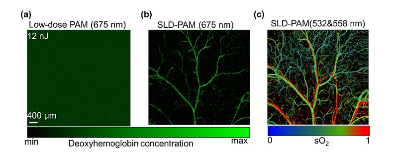
Comparison of the in vivo results of (a) low-dose PAM and (b) SLD-PAM, both at super-low pulse energy with a red-light source, and (c) oxygen saturation image acquired by SLD-PAM via dual-wavelength spectrum unmixing. Courtesy of Zhang, Y. et al., https://onlinelibrary.wiley.com/doi/10.1002/advs.202302486.
SLD-PAM can be used with a broad range of low-absorbing nano-agents, small molecules, and genetically encoded biomarkers, as well as low-power light sources in a wide spectrum. The system’s cost is relatively low, making it affordable for research laboratories and clinics.
“SLD-PAM enables noninvasive imaging of biological tissue with minimal damage to the subjects, offering a powerful and promising tool for anatomical, functional, and molecular imaging,” Wang said. “We believe that SLD-PAM can help advance the applications of photoacoustic imaging, enable numerous new biomedical applications, and pave a new avenue for clinical translation.”
Wang and his team plan to test a broader range of small molecules and genetically encoded biomarkers in biological imaging using the SLD-PAM system. They also intend to adopt more types of low-power light sources in a wider spectrum to develop wearable or portable optical resolution photoacoustic microscopy systems.
The research was published in Advanced Science (www.doi.org/10.1002/advs.202302486).
Published: September 2023