
Bioimaging Software Updates
MBF BioscienceRequest Info
WILLISTON, Vt., March 25, 2008 -- MBF Bioscience said it has released new, enhanced versions of its Stereo Investigator and Neurolucida quantitative imaging software packages and has also updated AutoNeuron, Neurolucida's advanced module for automated neuron reconstruction.
The new 8.0 version of Stereo Investigator for unbiased stereology handles the widest spectrum of researchers' needs and now has enhanced support for confocal, multichannel fluorescence and bright-field microscopy, MBF said. New workflow technology and probe interfaces assist with stereology, allowing researchers to be more productive and to obtain efficient, precise, unbiased, and reliable quantification of the number and morphometric properties of biological structures.
The unattended data collection and offline stereology feature enables image capture for analysis away from the microscope, and new mobile licensing allows the software to be moved between multiple workstations. Designed for compatibility with 32-bit and 64-bit Microsoft Vista, version 8.0 enables researchers to load and work with larger images, image stacks, and virtual slides.
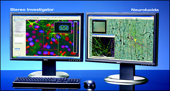 MBF Bioscience said it has added numerous enhancements to its Neurolucida software (used for 3-D neuron reconstructions) in its 8.0 version. Improved 3-D visualization, with the faster and smoother 3-D solids module, provides researchers with the ability to rotate and view image stacks in real time. Other enhancements include the ability to transfer results directly into Microsoft Excel. As with Stereo Investigator, the mobile licensing feature allows the software to be moved between multiple workstations, the company said.
The most advanced scientific software for performing brain mapping, neuron tracing, and morphometry, Neurolucida has the flexibility to handle data in many formats, MBF said. Researchers can use live images from digital or video cameras, acquired images from confocal microscopes, electron microscopes, and scanning tomographic sources; or through the microscope oculars using MBF's Lucivid system.
When combined with Neurolucida, AutoNeuron 3.0 performs automated neuron tracing in under a minute, and researchers can reconstruct neurons in 2-D and 3-D many times faster than manual tracing. Developed with the support of the National Institutes of Health, it rapidly explores the entire image volume and identifies neuronal processes and somas.
Version 3.0 introduces an enhanced workflow with improved detection and 3-D contouring of multiple somas, as well as advanced settings for branch connections and tracing guidance, the company said. AutoNeuron models neuronal trees as branching structures, complete with branch points, roots, and endings. Diameters of the axons and dendrites are automatically detected, and results can be transferred directly into an Excel spreadsheet.
For more information, visit: www.mbfbioscience.com; e-mail: [email protected]MBF Bioscience
185 Allen Brook Ln., Suite 101
Williston, VT 05495
Phone: (802) 288-9290
Fax: (802) 288-9002
https://www.mbfbioscience.com
/Buyers_Guide/MBF_Bioscience/c9158
Published: March 2008
REQUEST INFO ABOUT THIS PRODUCT
* First Name:
* Last Name:
* Email Address:
* Company:
* Country:
Message:
When you click "Send Request", we will record and send your personal contact information to MBF Bioscience by email so they may respond directly. You also agree that Photonics Media may contact you with information related to this inquiry, and that you have read and accept our
Privacy Policy and
Terms and Conditions of Use.
Register or login to auto-populate this form:
Login
Register
* Required