Anne L. Fischer, Senior Editor
Scientists study samples, such as microscopic detail of cellular tissue from a mouse kidney and live yeast cells, for obvious reasons. By examining these, they can begin to understand the inner workings of cellular systems, which could lead to the discovery of causes of and cures for human diseases.
A variety of microscopic techniques are available for such analysis, and researchers may not be aware of how images of the same sample can differ depending upon which one is employed. Wide-field fluorescence and spinning-disc confocal microscopy, for example, will yield different results because the latter blocks out-of-focus light during live-cell imaging.
Researchers can explore both conventional and emerging microscopic techniques at the Nikon Imaging Center, part of the cell biology department at Harvard Medical School in Boston. Jennifer Waters Shuler, director, helps guide graduate students and other researchers in the study of live cells and fixed tissues through various imaging techniques, as well as 3-D reconstruction of a sample based on several images taken at various depths.
Give and take
Its equipment distinguishes the center as one of the leading microscopy research facilities in the world, where scientists are introduced to the latest innovations in light microscopy and imaging. They gain insight into advanced techniques and, in turn, provide valuable feedback to equipment suppliers.
Workstations set up throughout the one-room facility are designed for different types of light microscopy. They use microscopes from Nikon Inc.’s Instruments Group of Melville, N.Y., supported by equipment on loan from or donated by corporate sponsors. Two stations have an inverted light microscope (model TE2000) with a PerkinElmer spinning-disc confocal. One also has a Solent Scientific incubation chamber, and another station has the same type of microscope with a laser scanning confocal. The first has a key strength in live-cell imaging; the second in 3-D reconstruction applications.
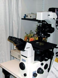
The Eclipse TE2000 inverted research microscope from Nikon Instruments allows multiple epifluorescence techniques to be used simultaneously or sequentially.
Also available are a total internal reflection fluorescence system; three additional upright microscopes, including one with a Bio-Rad laser scanning confocal; and an inverted fluorescence microscope (TE2000E) with Eppendorf micro-injection. Support equipment includes Hamamatsu CCD cameras, Chroma Technology filters and heated chambers for live-cell research. There are also computers at each workstation that have Metamorph image analysis software, AutoDeBlur deconvolution software and AutoVisualize 3-D rendering software.
The inverted light microscopes used at two stations feature a multiport design and a stratum structure that provide configuration flexibility. These microscopes are prevalent in the lab because they can use multiple optical accessories either separately or simultaneously, supporting multiple input illumination sources as well as output detector functions. Multiple epifluorescence techniques may be easily used simultaneously or sequentially. Systems thus allow other techniques such as high-resolution deconvolution of 3-D images, which could be incorporated with fluorescence recovering after photobleaching applications.
Laser illumination techniques also include optical tweezers and laser microdissection. A stray-light noise terminator captures images with unprecedented signal-to-noise ratios. With the inverted light microscopes and a dedicated communications hub controller, researchers can choose retrofittable motorized options to fine-tune their methods. It also is possible to control the microscope externally from a connected PC, making it easy to integrate image analysis using commercially available image processing software.
Point and shoot
To ensure greater stability, Nikon adopted a new, high-strength alloy material, M-45, in the microscope base that doubles the rigidity and improves the precision of the focusing movement, as compared with previous models. Because the thermal stability is as important as rigid construction, the company also designed the main body structure so that minor ambient temperature fluctuations do not adversely affect image capture. This design is particularly useful for time-intensive research such as time-lapse recording.
High-resolution imaging with research-grade equipment generally requires user attention that makes point-and-shoot microscopy difficult, but advances in hardware — such as in digital cameras and automated filter wheels and shutters — have simplified image acquisition, according to Waters Shuler. Although she doesn’t believe that research-grade light microscopy should even attempt to reach the level of point-and-shoot, she indicated that recent advances include prepackaged commercial systems for total internal reflection — turnkey sets that use various types of lasers at different wavelengths.
Beyond simplification of setup for microscopy, she would like to see cameras that are more sensitive and better, lower-cost lasers, including devices with more stability. In line with this, Nikon is working with various laser companies to integrate solid-state lasers into the total internal reflection and confocal sets.
Steve Ross, a senior applications scientist with the microscope manufacturer, believes that diode lasers are superior to gas lasers because of their longevity, steady output and range of wavelengths. He anticipates that the firm will introduce products next fall that are designed to advance laser-based microscopy.
Contact: Nikon Imaging Center, Boston; +1 (800) 645-6687; Web: www.nikonusa.com.