John P. Murphy, PerkinElmer Inc.
Digital flat panel x-ray detectors illustrate how an established technology can penetrate a new, high-growth market. This is apparent in their use in medical imaging, where they continue to show great promise for growth as a result of their diagnostic capabilities.
Digital x-ray detectors continue to show growth.
Two major suppliers of medical imaging products, GE Healthcare in Chalfont St. Giles, UK, and Elekta AB in Stockholm, Sweden, have announced that they plan to use the detectors for advanced medical imaging in the coming years. Because digital x-ray technology increases the medical professional’s ability to capture, store and retrieve images and because it improves the diagnosis and treatment of patients, it is no wonder that the estimated growth rate for the digital flat panel x-ray detector market worldwide is $250 million and growing at 20 percent per year.
Optoelectronics companies are becoming increasingly market-focused, developing custom solutions for particular industries, such as automotive and consumer electronics. In the case of the medical imaging industry, technology-driven companies are recognizing that an awareness of new products and secondary application opportunities can give them a competitive edge.
With this perspective, medical imaging has become a focal point for flat panel digital applications, replacing traditional film radiography in hospitals and improving accuracy, efficiency and productivity. For example, hospital technicians can position a patient, take an image and review it in seconds, knowing immediately whether the image is valid or requires a retake. The technology completes in minutes processes that previously might have taken hours or even days.
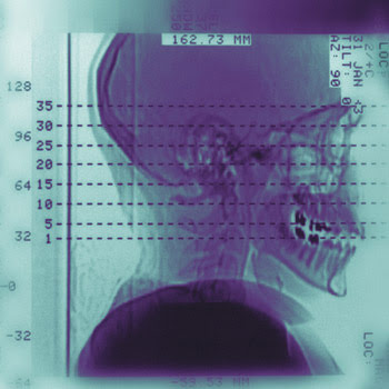
High-performance digital x-ray technology is based on amorphous silicon (a-Si) fabricated on a glass substrate using thin-film processing. An x-ray scintillator, which converts x-rays to visible photons, is grown directly on or is attached to the a-Si panel. These photons are converted into an electrical charge that is, in turn, converted into a digital value for each of several million addressable pixels on the panel to create the image.
Hospitals typically can replace two film rooms used in traditional radiography with a single digital unit. Other benefits include the elimination of processing chemicals and the reduction of space, including the need for large x-ray storage rooms.
A sister application within the medical environment is real-time imaging for diagnosis in cardiology or angiography. For this application, flat panel digital detectors are replacing traditional image intensifiers. Physicians can view real-time blood flow in the body at 30 images per second. By injecting a contrast agent, or x-ray-absorbing dye, into the patient’s vascular system, the blood flow can be viewed in real time to locate and alleviate blockages. The image from the detector is much sharper than those created with image intensifiers.
Digital subtraction angiography — subtracting an image taken without a contrast agent from an image taken with one — also is more easily achieved. The subtraction process further highlights the vascular system by removing other elements, such as bone, from the image. However, this process might be eliminated as a result of the flat panel digital detector’s ability to achieve sufficient visibility in just one image using a contrast agent. This not only provides a work-flow benefit, but also lowers the x-ray dose to the patient.
Digital flat panels also provide patient imaging for radiotherapy systems. Online image acquisition controls the patient’s position during a cancer treatment. Because they distinguish even the smallest contrast differences in bone and tissue, these detectors can provide treatment at lower applied doses. Their high frame rates also will enable enhanced treatment methods, such as intensity-modulated radiotherapy.
In diagnostic medical applications, a 1000 × 1000-pixel image at 30 images per second can be achieved. There are detectors available that can produce a 2000 × 2000-pixel image, although some speed is traded for image size. Future applications may require 3-D imaging at the same high speeds, and products that can satisfy such applications are on the horizon. Also, as costs begin to drop for flat panel digital technology, lower-priced, portable x-ray systems may be wheeled from room to room in medical centers, saving time and expense.
Applications for these detectors have increased, and their use in today’s medical imaging applications continues to grow rapidly. Such growth likely will continue to drive the next generation of medical imaging diagnostics.
Meet the author
John P. Murphy is executive vice president and chief operating officer at PerkinElmer Inc. in Boston; e-mail: [email protected].