A solid-state x-ray image intensifier is used for diagnostic and interventional medical imaging procedures.
Andrew Kuhls-Gilcrist and Stephen Rudin, Toshiba Stroke Research Center
With the onset of improved, patient-specific treatments for vascular disease, along with advancing diagnostic techniques, there is an increasing need for high-resolution images obtainable in real time. Current state-of-the-art medical x-ray image intensifiers have inherent limitations and are being replaced with flat panel detectors.
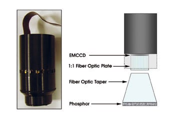
The solid-state x-ray image intensifier prototype consists of a modular array of electron-multiplying CCD camera systems that are modified to image x-rays with an increased field of view (left). Interchangeable components enable task-based optimization of the detector system for both diagnostic and interventional medical imaging procedures.
Flat panel detectors are used for dynamic x-ray imaging in fluoroscopy and angiography because they do not suffer from veiling glare or from pincushion or S-shaped distortion. However, their performance has been disappointing for low-exposure-per-frame procedures because they suffer from excessive instrumentation noise, lag and ghosting. Additionally, both detectors have resolution capabilities of only roughly three line pairs per millimeter.
To overcome these limitations, the Toshiba Stroke Research Center at the University at Buffalo in New York has been developing a digital dynamic x-ray image receptor — designated the solid-state x-ray image intensifier — that has the advantages of flat panel detectors but with no lag or ghosting, and it has negligible instrumentation noise.

Shown are two comparative images taken with a state-of-the-art x-ray image intensifier (XII) and a recently developed solid-state x-ray image intensifier (SSXII) of an asymmetric stent used for treatment of neurovascular aneurysms. Both images were acquired using the same exposure condition and imaging geometry.
The prototype module is composed of an electron-multiplying CCD camera system manufactured by Photonic Science Ltd. of East Sussex, UK. It images a CsI(Tl) scintillating phosphor via a fiber optic taper. The camera’s sensor operates like a standard frame-transfer CCD, with the addition of a unique multiplication register before the readout of the signal. This built-in adjustable gain amplifies the signal, in solid-state, allowing for the effective elimination of read-noise degradations at lower x-ray exposures — important for minimizing potentially harmful radiation to the patient. A Peltier cooler reduces thermal electron noise in the sensor to negligible levels. Easily interchangeable front-end components enable task-based optimization of the detector system for both diagnostic and interventional medical imaging procedures.
Initial use of the solid-state x-ray image intensifier has hinted that the technology has great promise. Inherent detector resolution measurements have indicated the ability to resolve upward of 20 line pairs per millimeter. Images taken across a range of clinically relevant exposure conditions have demonstrated quantum-limited, instrumentation-noise-free images over more than three orders of magnitude, below which the incident photon statistics are so poor that a useful image no longer can be formed. Comparative images taken with a state-of-the-art x-ray image intensifier and the new solid-state x-ray image intensifier, under the same exposure conditions and imaging geometry, reveal that the solid-state imager can provide a substantial improvement over current technologies.
The next challenge is to increase the detector’s field of view. A 6:1 minifying fiber optic taper provides a field size of 4.8 cm per module. Work is under way to construct a 2 × 2 array of such modules for region-of-interest and neurovascular imaging. Larger arrays may be constructed to satisfy both cardiac imaging and general fluoroscopic applications. Module alignment, digital stitching and distortion-correction issues resulting from the array design are being addressed.
As electron-multiplying CCD technology matures, it is expected that the sensor cost will decrease. Sensors as large as 13 mm already are commercially available, which would facilitate the need for fewer modules. This would tend to further increase the cost-effectiveness of the new solid-state image intensifiers, while allowing them to be used in the same applications as current flat panel detectors.
Meet the authors
Stephen Rudin, PhD, is co-director of the Toshiba Stroke Research Center in Buffalo, N.Y.; e-mail: [email protected].
Andrew Kuhls-Gilcrist is a research assistant working in professor Stephen Rudin’s medical imaging physics group; e-mail: [email protected].