
Novel autofocus technique advances high-resolution, time-lapse imaging
Gary Boas
Development and continued improvement of fluorescent biosensors have provided researchers with access to intracellular protein dynamics in individual living cells — through high-resolution, time-lapse imaging of multiple fields of view, for example. Autofocus often is called on in these studies to compensate for temperature drift, for an uneven substrate across the multiple fields of view and for cell growth over extended periods.
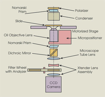
Researchers have reported a novel autofocus technique for high-resolution time-lapse imaging. The technique takes advantage of a specially designed bandpass finite impulse response filter to achieve maximal autofocus performance. Based on an inverted microscope, the autofocus module also includes an Extender lens assembly so that the optics in the filter wheel do not affect image formation at the CCD.
Designing high-precision autofocus systems is never simple, but it can be even more difficult when using the high-numerical-aperture oil-immersion objectives needed for high spatial resolution. In the July issue of Cytometry Part A, a team of researchers, three at the University of North Carolina at Chapel Hill and the other at Burnham Institute for Medical Research in La Jolla, Calif., and at Vala Sciences Inc. in San Diego, described how they addressed these challenges with a new autofocus technique for high-resolution oil-immersion microscopy using transmitted light differential interference contrast (DIC) imaging.
Biosensing revolution
The work was motivated in part by the biosensing needs of the researchers. The development of biosensors with very high spatial resolution is “creating a revolution in biosensing,” said Klaus M. Hahn, who, together with Jeffrey H. Price, was one of the two co-principal investigators of the study, “but you don’t see much of it in high-content screening, where the emphasis is on throughput.” Spatial resolution and sensitivity are typically sacrificed in high-content screening. “We are designing these biosensors with the goal of developing at least a medium-throughput system.”
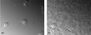
The investigators demonstrated a precision of 10.3 nm with the autofocus technique. In addition, using specimens with a range of thicknesses — 9.47 (a) to 33.20 μm (b) — they showed that the precision of the technique is independent of thickness. Reprinted with permission of Cytometry Part A.
They developed the technique for DIC imaging, rather than for fluorescence imaging. They chose the former for several reasons: to circumvent meniscus distorting phase contrast in microtiter wells, to avoid losing any fluorescence emission light resulting from the objective phase ring, and, finally, to avail themselves of the higher spatial resolution afforded by differential interference contrast, with respect to phase contrast.
They achieved maximal autofocus performance for DIC imaging by designing a new bandpass finite impulse response filter, which determines the spatial frequency band with the highest signal-to-noise ratio and the smallest distortion side peaks. Studies have shown that side peaks increase with the inclusion of additional lower frequencies in the passband. As a result, selecting the most appropriate spatial frequency band involves something of a trade-off: Excluding the lower frequencies increases the sharpness of the focus measurement maximum and, thus, improves the precision but results in low signal-to-noise. Including them, on the other hand, improves the signal-to-noise. The researchers designed the new bandpass filter with this in mind.
They needed to address various mechanical issues as well. For example, a sort of spring tension develops in the oil that will drive the objective back, “so we had to change some of the movements that the objective made,” Hahn said.
They assessed the performance of the new technique in a series of autofocus trials. The experimental setup consisted of an image cytometer based on an Olympus microscope with a 60×, 1.4-NA oil-immersion objective, with the digital autofocus module built onto it. Seeking to increase the contrast and improve the autofocus performance, the researchers removed the analyzer from the fluorescent light path in the microscope. A 12-bit monochrome scientific-grade digital Photometrics CCD camera interfaced with the computer via a PCI card captured images for autofocus. This combination facilitated fast region-of-interest acquisition, enabling them to focus on a specified 600 × 600-pixel region in the center of the field of view.
Tests on 225 fixed-cell samples revealed a precision of 10.3 nm; further trials with specimens of varying thickness — from 9.47 to 33.20 μm — confirmed that the autofocus precision was independent of thickness. The results showed that, by selecting particular spatial frequencies, they could achieve very high precision autofocus for high-numerical-aperture DIC automated microscopy — enabling stable focus for high-resolution time-lapse imaging even over extended periods and helping to overcome the issues typically associated with such studies.
A range of applications could benefit from the new autofocus technique. “There’s a huge realm of signaling that depends on determining the precise timing and localization of protein activation,” Hahn said. This includes studies of cell movement and plasticity as well as rare cell analysis. For example, if investigators were to find that a drug is effective in some areas of a particular cell population — say, a tumor biopsy — but not in others, they might want to analyze the drug impact on specific cells.
They plan to incorporate the autofocus technique into their research in several ways. “Primarily, we’re going to be looking at metastasis,” Hahn said, “where you need to understand the proteins that control coordination of signals with submicron and subsecond resolution.”
Contact: Klaus M. Hahn, University of North Carolina at Chapel Hill; e-mail: khahn@med.unc.edu; Jeffrey H. Price, Burnham Institute for Medical Research; e-mail: jprice@burnham.org.
Published: September 2008