
Retinal OCT Comes of Age
A new generation of OCT instruments ups the ante in ophthalmology with fast image acquisition and higher-resolution 3-D volumetric imaging.
Gary Boas, News Editor
The emergence of optical coherence tomography (OCT) in the past decade and a half has yielded tremendous benefits for the field of ophthalmology. By producing cross-sectional images of the eye based on depthwise reflections of near-IR light, the technology has provided a relatively quick, inexpensive and user-friendly means for diagnosing particular eye conditions and for monitoring retinal health.
Now, thanks to advances in detectors and light sources, a new wave of instruments is offering improvements in both speed and resolution, allowing 3-D volumetric imaging and contributing to treatment and monitoring of a range of conditions.
A new generation of ophthalmologic OCT instruments uses spectral rather than time-domain technology, producing much faster scans and even enabling 3-D volumetric measurements.
In a 1991 Science paper, the concept of OCT was introduced by James G. Fujimoto’s group at MIT in Cambridge, Mass. With David Huang as the primary author, the paper also demonstrated the first cross-sectional retinal images. OCT’s antecedents lie in optical time-domain reflectometry and optical coherence-domain reflectometry, which the telecommunications industry developed to identify the locations of faults in fibers. With these techniques, operators launched optical pulses into the fibers. If they detected any reflections, they knew they had found a fault. Furthermore, based on the time delay, they could determine where — that is, how far down the fiber — the fault was located.
OCT is based on the same principle. Instead of taking only a single depth scan, though, the technique draws on data from adjacent positions in tissue. “It represents the assembly of those data to form an image,” said Dr. Stephen A. Boppart, who joined Fujimoto’s group in 1993 and currently heads the Biophotonics Imaging Laboratory at the University of Illinois at Urbana-Champaign.
Because OCT provides high-resolution cross-sectional images through the retina, it allows ophthalmologists to see conditions such as a detached retina, macular edema and macular holes, and even changes in the retinal nerve fiber layer.
Indeed, investigators found that OCT’s high-resolution images of subsurface retinal features that were inaccessible to previous gold standard techniques — fluorescein angiography and ophthalmic ultrasound — were uncovering a variety of previously unknown features of ocular disease. For example, researchers ended up creating a new category for staging macular holes. The staging in its previous form had been used for years, but researchers realized that events transpire prior to Stage 1.
MIT licensed the original OCT technology to Humphrey Systems — now Carl Zeiss Meditec Inc. of Dublin, Calif. — who commercially developed it specifically for ophthalmic applications. The initial instrument was introduced in 1995, followed by the smaller, more user-friendly OCT2 in 2000, and then by the Stratus OCT in 2002. The Stratus was smaller and faster still, and it offered much higher resolution than either of its predecessors.
The latter instrument, especially, was embraced by the ophthalmology community. Carl Zeiss Meditec has sold nearly 9000 since its introduction, said the company’s Marianne Whitby, with roughly half of those sales in the US. The primary applications of OCT have involved scanning the retina to detect the presence of, or any change in, retinal disease, or to diagnose glaucoma. The technology has a broader reach in the field, however. “Even doctors who aren’t dealing with retinal disease or glaucoma want to know the retinal health of the eye,” Whitby said.
In New York City, an optometric group practice comprising Dr. Arthur Jung, Dr. Frederic Nevins and Dr. Traci Goldstein uses the instrument, which it acquired in early 2005, routinely. “When patients have very subtle complaints of vision loss or, as a doctor, we are unsure why a patient’s visual acuity is decreased, a fast macular or retinal nerve fiber scan can reveal a problem that can be attended to,” Jung said.
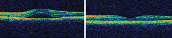
Using OCT, ophthalmologists in New York diagnosed central serous retinopathy in a patient who had noticed a blue haze over one eye. Shown here are OCT images revealing the condition (left) and demonstrating the normal macula (right).
In one such case, a patient had noticed a slight blue haze over one eye. His vision was 20/20, and all other findings were normal. The OCT helped make the diagnosis very easily: The patient had a condition called central serous retinopathy, in which fluid accumulates underneath the retina and thus causes visual distortions and even vision loss. Without the OCT, he would have had to undergo a fluorescein angiogram, a procedure during which a dye is injected into the bloodstream and images are acquired to see whether the dye leaks into the retina. “This is the standard of care but is more time-consuming and can be stressful to the patient,” Jung said.
Whitby noted another possible application. As more therapies become available for age-related eye disease — age-related macular degeneration, for example — doctors are increasingly interested in identifying and monitoring the earliest stages of the disease so that they can address it appropriately, and they want to watch the retina so that they can time the treatment properly.
The next generation
In the past couple of years, a range of technological advances has produced faster OCT systems with much higher resolution than previously was possible. These advances were made possible with Fourier-domain detection techniques such as spectral-domain OCT and swept-source OCT, which themselves were the outcome of advances in detector and light source technologies.
The improved acquisition rates have yielded another important development: 3-D volumetric imaging. In 2007, Carl Zeiss Meditec introduced the Cirrus HD-OCT, which uses spectral-domain rather than time-domain technology. The instrument scans much faster as a result and thus can provide 3-D volumetric measurements: Instead of offering single cross sections, it assembles 128 or 200 cross sections to create a cube of data.
The much faster acquisition rate of Fourier-domain OCT — 26,000 axial scans per second, as compared with the 400 axial scans per second offered by the Stratus OCT — can aid optometrists and ophthalmologists in other ways: helping to avoid issues related to subtle eye movements and blinks, for instance. The acquisition rate and resolution of the earlier instrument already may be high enough for many applications, but other applications could benefit from the increased speeds, Jung said. “In clinical practice, the Stratus has been shown to provide us with enough information to diagnose and monitor most retinal conditions and glaucoma. But the newer OCTs show much promise, and I’m sure we will upgrade in the near future.”
The advances have produced whole new sets of concerns, though. Whereas in the early years the method was limited mostly by the optics — how can we scan fast enough? — the challenges now fall primarily in the areas of signal and image analysis. “It’s been an interesting shift over the years,” Boppart said. “We’re able to scan so fast now that we’re limited by processing power and how fast we can manage the data. The onus is on the computer industry to catch up.”
The corporate and patent landscapes have been changing, as well. As the technology has evolved and diversified, new patents have been filed, and a number of new players have entered the market. Carl Zeiss Meditec still rules the roost, holding more than 10 of the 40 US patents with OCT in the title as of September 2007, when Boppart and colleagues published a review of the field in Journal of Biomedical Optics. The field is changing rapidly, however, as new companies join the fray. A handful of these are focusing on high-speed retinal imaging, including Optovue Inc. of Fremont, Calif.; Topcon Corp. of Tokyo; Optopol Technology SA of Zawiercie, Poland; Heidelberg Engineering Inc. of Heidelberg, Germany; and Ophthalmic Technologies Inc. of Toronto.
The market has continued to grow since these others first appeared on the scene, in about 2006. By the company’s estimation, Carl Zeiss Meditec still commands about 30 percent of the market, selling approximately 1600 Cirrus instruments in the past year. Whitby notes, though, that the overall growth of the market since 2006 is likely because of the emergence of the new players.
OCT-Guided Tissue Cutting
Optical coherence tomography can contribute to ophthalmic applications even beyond diagnosis and treatment monitoring. The Hannover, Germany-based company Rowiak GmbH has designed an instrument that combines OCT and laser microtomy to enable OCT-controlled cutting of biological tissue — facilitating slicing as well as ablating inside the tissue. "This is, in our view, the first step toward 3-D laser microdissectionm" said the company's Fabian Will, who was responsible for development of the system.
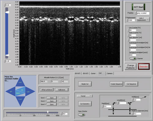
A novel instrument combines OCT and laser microtomy to offer 3-D monitoring and guidance of tissue cutting procedures. The instrument can aid in a variety of ophthalmologic applications.
Tissue cutting is performed using a femtosecond laser, which a high-numerical-aperture objective tightly focuses into the sample. The high intensities inside the focal volume lead to nonlinear absorption processes and finally to disruption of the material within the focal diameter of about 1 µm. The instrument moves the laser beam and the sample simultaneously during cutting, the laser beam using a fast scanner and the sample using a piezo-driven positioning stage.
The instrument performs 3-D imaging and guidance of the cutting process by using a modified Thorlabs Spectral Radar 2-D OCT system with an additional OCT module containing the reference arm and optics for dispersion compensation.
The instrument, which was developed with Laser Zentrum Hannover, facilitates a range of applications, including cutting along predefined cell layers, cutting out volumes of cells for further cultivation, and sectioning of tissue at predefined structures. Possible uses within ophthalmology
include optimization of flap thickness for lasik — enabling doctors to get as close as they are able to Bowman's membrane without damaging it — and optimization of cornea transplants of the endothelium.
In collaboration with Hannover Medical School, the company has demonstrated the potential of the instrument by performing OCT-guided cutting in porcine and human corneas along defined cell layers as well as in small volumes of porcine skin and cornea samples.
Published: September 2008