Which imaging modality is right for your small-animal research?
Richard Gaughan, Contributing Editor
In light of rising health care costs, technology that improves the efficiency of medical research is welcome. Small-animal imagers promise to streamline therapy development by imaging biological processes in mouse or rat models of human disease. They offer a variety of individual and combination imaging modalities, including fluorescence, bioluminescence, x-ray (CT), PET, single-photon-emission computed tomography (SPECT), MRI and ultrasound – just about any clinical imaging modality is available in a small-animal instrument.
With a wide array of imaging techniques, what governs the choice of modality? Todd Peterson, assistant professor in the department of radiology and radiological sciences at Vanderbilt University in Nashville, Tenn., notes that “Often, people try to stretch a specific modality to measure just about everything.” But each modality has its advantages and disadvantages.
Light and easy
For ease of use and short acquisition time, optical imagers are the prime selection. Yingqiu Yvette Liu, technical director of the Small Animal Imaging Facility at the University of Pennsylvania in Philadelphia, said, “A bioluminescence image can be acquired in a minute or two, and fluorescence images can take just seconds.”
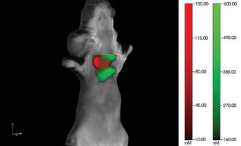
Traditional optical systems are planar imagers that project all the received photons onto a single plane. VisEn Medical offers a fluorescence molecular tomography system that reconstructs a three-dimensional representation of the source emission. Transilluminating a mouse and collecting light over a range of angles enable accurate imaging of organs deep within the body. Here a mouse model of pulmonary inflammation is labeled with two distinct dyes to separate pulmonary edema from neutrophil influx. Courtesy of Dr. Jeffrey D. Peterson at VisEn Medical.
Bioluminescent model organisms typically express luciferase when a protein of interest is expressed, allowing researchers to visualize gene expression. The technique requires injection of luciferin substrate; then luciferase will emit light in 10 to 25 minutes. The weak bioluminescence signal requires a sensitive detector. The first commercially available bioluminescence imager was from Xenogen (now part of Caliper Life Sciences). The company’s current IVIS instrument gets its sensitivity from a 1-in.-square CCD chip cooled to 290 °C.
Fluorescence imaging relies on the expression of fluorescent proteins or injected fluorophore-bearing molecules that bind to specific target molecules. According to Bradley Smith, director of Notre Dame’s Integrated Imaging Facility in Indiana, optical imaging is familiar to biological researchers because a lot of cell biology studies use fluorescent probes. “You can pretty much take the same technology [as used in cell biology], extend it to longer wavelength emission, then squirt it into an animal and see it,” Smith said.

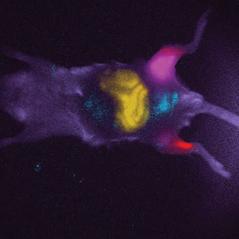
With current commercially available instruments, optical imaging is relatively straightforward and easy, yet it remains a powerful technique. Whether imaging bioluminescent activity within a mouse or GFP fluorescence in a genetically engineered mouse, a single image can reveal layers of biological information. Images courtesy of Yingqiu Yvette Liu.
Notre Dame is acquiring a Kodak multispectral system from Carestream Molecular Imaging of New Haven, Conn., and Smith plans to make use of its spectral unmixing capability. This capability can separate a probe targeting inflammation from another specific to bacterial infection, which could allow Smith to study how long it takes after bacterial infection for the inflammatory response to begin and, when evaluating drug response, to study the timing of the clearing of the bacteria and to quantify the relaxation of the inflammatory response.
Fluorescent dyes come in many colors, but they are not necessarily clearly distinguished in the in vivo imaging environment. Instruments like the Kodak in vivo multispectral imaging system FX from Carestream Molecular Imaging use excitation and emission filters to identify signals from the various fluorophores and to separate them from autofluorescence.
But for all its ease and utility, imaging is limited by scatter and absorption in living tissue. Christopher Flask, scientific director of Case Western Reserve University’s Small Animal Imaging Center in Cleveland, put it succinctly: “If you’re looking for something subtle, optical inside is probably not the way to go.”
Cherchez l’eau
“Of the variety of applications that come in to the center, the majority have an MR component to them,” Flask said. “MRI has really great soft-tissue contrast. There’s great value in just being able to put the animal on the table and 10 minutes later ‘see’ the organs.”
MRI produces images of water by exciting the magnetic moment of the single protons that form the nucleus of hydrogen atoms. The excited protons return to their equilibrium state, emitting radio-frequency signals as they do. The temporal characteristics of the emitted radio frequency provide a signature for the surrounding tissue.
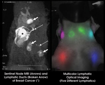
MRI and fluorescence imaging capabilities are complementary. MRI provides anatomical detail even deep within the mouse, while fluorescence provides an identifiable signal for various molecular markers. These two views of the lymphatic system illustrate the differences. Courtesy of Hisataka Kobayashi.
Imaging small animals with MRI has revealed some important information about diseases that cannot be revealed simply by dissecting a mouse model. One example, Flask said, is vaginal prolapse, a condition that can occur later in life when the network of muscles, ligaments and skin around a woman’s vagina weaken or break. Dissecting the mouse model of this condition couldn’t answer simple questions because tissue would move. “Then, 15 minutes of scanning changed the whole impression of the disease,” Flask said. The MRI produced by the center’s Bruker Biospin 9.4-T MR scanner revealed that bladders of mouse models had been anatomically displaced to slip down below the symphysis bone.
MRI’s intrinsic spatial resolution can get down to a couple hundred microns but, according to Mark Does, director of the Center for Small Animal Imaging at Vanderbilt University, it’s possible to infer tissue characteristics at much smaller scales because diffusion-weighted MRI impresses a spatial order on the magnetic moments. If that order persists, then a large signal is returned; if motion has reduced the order, the signal diminishes. When the water motion is hindered or restricted by membranes or other microanatomy, the apparent diffusion constant (ADC) is reduced. “For example, water ADC is higher in the direction of axons than in the direction perpendicular, so axonal orientation can be measured,” Does said.
Generally, MRI offers the best soft-tissue imaging, but it doesn’t provide molecular specificity because the user must infer the molecular and chemical situation based on the effect on water, Does said. Strategies for molecular-specific MRI contrast have been explored, but for studies in which it is necessary to identify particular cells or specific molecular levels, he said, “MRI probably isn’t the optimal choice.” For specificity and good spatial resolution, radioisotopic imaging may just fit the bill.
What the doctor ordered
SPECT and PET image the decay products of radioactive isotopes incorporated into marker molecules. “SPECT and PET have the sensitivity: They get past the absorption issue,” Flask said. SPECT images the gamma radiation resulting from the decay of isotopes, typically technetium or iodine, and PET images the pair of gamma rays generated when an electron is struck by a positron coming from the decay of an isotope of an atom such as fluorine or carbon. Often, a glucose molecule is modified with the addition of a radiolabel to track metabolic activity.
Radioisotopes decay and either directly or indirectly emit gamma rays. Bioscan’s NanoSPECT instrument reconstructs the gamma ray sources with resolution as good as 0.4 mm. Tying those radioisotopic images to structural images from the companion CT imager enables clear and unambiguous identification of the source organs.
The factors that determine the choice between PET and SPECT, according to Peterson, often are practical rather than technological. For example, “SPECT has a wider range of radiotracer kits already labeled, while for PET you need nearby access to a cyclotron and then the synthetic chemistry to assemble the complete radiolabel – it’s still a fair bit of work even for standard radiotracers.” Timing is also important. For example, antibody labels take a while to circulate through the body, so those studies typically want to image over the course of days. The balance between the uptake and clearance peaks is often optimum after about a day, but if that’s equivalent to several half-lives for the radiolabel, a lot of the imaging capability is gone.
Peterson is now designing an experiment to equivalently compare SPECT and PET imaging. One potential experiment would evaluate imaging of osteolytic bone lesions resulting from metastases. “A lot of the value of in vivo imaging comes from the ability to do longitudinal studies in the same organism. For long-term studies, dose is a concern,” he said.
For example, in a situation where one method provides more sensitivity than another, one would assume that the difference could be compensated for quite simply: Just up the dosage of the radiolabel. However, this seemingly obvious solution overlooks the important fact that total dose is important – especially for long-term studies. So to truly compare the techniques on equal terms, the total dosage must be the same.
Radioisotopic imaging provides a reasonable-resolution, relatively background-free, high-sensitivity image of radiolabel concentration. But relating that signal to specific anatomical regions is not always unambiguous. The images are more valuable if the signal’s location can be identified with confidence. Multimodal imagers can help achieve this.
Notre Dame’s Smith said that newer capabilities, such as coregistration of fluorescence with CT, help add spatial location. “It adds detail to the typically ‘blobby’ fluorescence signal to be able to definitely state things like ‘that is the kidney,’” he said.
Case Western’s Flask noted that radioisotopic imaging, joined with CT and instant overlay capability, reconstructs the real source and couples it to anatomy. Vanderbilt’s Does agreed that it makes sense to go multimodality because it cuts down on moving the animals. Although CT is the common companion modality right now, PET/MRI combined instruments are coming, Does said. He warned, however, that issues with coregistering data are not trivial.
Regardless of the imaging modality, a key component of image quality – and diagnostic utility – is contrast. No matter the modality, more contrast is better.
Hisataka Kobayashi, chief scientist in the molecular imaging program at the Center for Cancer Research at the National Cancer Institute in Bethesda, Md., develops passively or actively target-specific contrast agents. These include contrast enhancement agents for SPECT, PET, ultrasound, MRI and x-ray imaging as well as fluorescent markers for optical imaging. First he identifies agents that work well in vitro. “Maybe less than one-tenth of the contrast agents that show promise in vitro,” he said, “will also demonstrate favorable binding characteristics in vivo.”
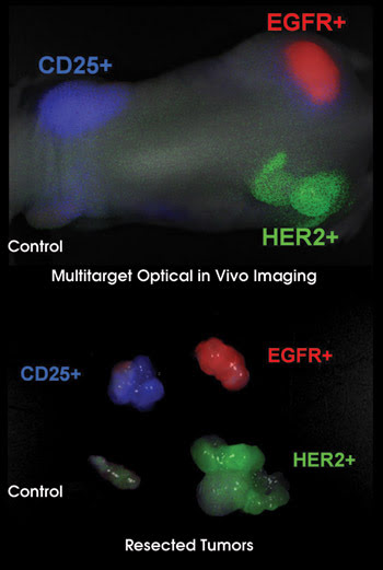
By linking fluorophores with molecular antibodies, specific molecular targets can be imaged in vivo. Here three fluorescent probes identify tumors expressing different molecules. The in vivo image is confirmed by fluorescent imaging of the explanted tumors. Courtesy of Hisataka Kobayashi.
Small-animal imagers allow more efficient and more accurate measurements of diagnostic in vivo performance of contrast agents. Longitudinal studies of the same animals require fewer sacrifices, and correlating images over time in the same mouse improves confidence in the measurements, no matter what the modality. To determine the appropriate imaging modality, he considers the expression level of the target molecule, the depth of the target organ/object and the required spatial resolution. “The modality to use,” he said, “is the combination of modality and contrast agent that will meet my goal.”
Are you new to small-animal imaging?
In vivo small-animal imaging offers unique advantages. It is minimally or entirely non-invasive, examines biological processes in their native environments, uses clinical modalities and allows longitudinal studies. Those compelling advantages are driving medical and biological researchers to consider incorporating small-animal imagers into their research protocols. What is most important for them to keep in mind?
Bradley Smith suggested a first conversation might revolve around the selection of a labeling method. The experimenter must choose between ligand-bound probes that stick to the target and the activated probes that emit, for example, when they’re presented with their complementary enzyme.
Mark Does noted that new users are used to thinking in terms of what they can look at with a confocal microscope. “You can’t possibly get the same level of detail in vivo as you can observing excised tissue on a slide.” New users often don’t appreciate that in vivo imaging is generally on the macroscopic scale, and the challenge is to relate macroscopic contrast to microscopic characteristics.
For Christopher Flask the issue involves “who” as much as “how.” Often the expectations of the biologists are not realistic. “Cultivating imaging experts is good.” He suggests consulting with an MR physicist, an optical imaging expert or whoever is appropriate to convey the capabilities of the imaging modalities in question.
But even with the limitations and the experimental protocol design concerns, Yingqiu Yvette Liu summarized the promise of small-animal imaging: “The trend toward translational research is designed to enable a lot of applications to quickly move from the preclinical to the clinical stage, and many applications are only a small step away from achieving this transition.”