 A team of dentists and physicists in the UK is developing an infrared imaging system for dentistry that avoids the risks associated with x-rays. Professor John Girkin and Dr. Simon Poland at the University of Strathclyde’s Institute of Photonics in Glasgow, Scotland, have worked with dental health specialists professor Nigel Pitts and Dr. Chris Longbottom of the University of Dundee to develop the method. It allows dentists to examine the interior of every tooth at every visit, enabling less invasive treatment to begin at a much earlier stage. It also facilitates close monitoring of complex procedures.
Risks from ionizing radiation limit the number of x-rays dentists are permitted to take, so they usually must examine the tooth by eye. But dental caries develop just below the surface of the tooth with mineral loss occurring at increasing depths, and in the crucial initial stages when the process is reversible, the tooth appears undamaged until the problem is quite advanced. Under normal white light, early lesions show up as whiter areas against the white background of the surrounding tooth material, making them hard to spot against the shiny and inaccessible surfaces within the mouth.
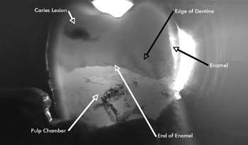
This infrared transmission image shows the internal structure of a tooth. Courtesy of University of Strathclyde, Glasgow, UK.
The infrared imaging system is based on a near-infrared laser diode of the type used in CD players; one version uses low-cost CCD cameras. The beam shines through the tooth, and the camera builds an image that is the optical equivalent of a conventional x-ray. Because infrared light is nonionizing, it poses no risk to the patient or the dentist, allowing frequent examinations to monitor the progress of a lesion. The method provides images of a quality similar to that of x-rays and can provide real-time images of the tooth and root within the jaw, making the system ideal for monitoring pediatric orthodontic and endodontic treatments.
A variation of the basic design uses optical components present in a digital light projector to direct the light off the tooth, enhancing the contrast between healthy and demineralized tissue. With no lead shielding, photographic plates or chemical processing, the capital and running costs of the infrared system are lower than the price of x-rays.
The development won three prizes in December 2008 at the Medical Futures Innovation Awards in London, attracting much business support. The team is testing the device and looking for funding for commercialization, via company start-up or technology licensing to interested parties around the world. Professor Girkin has very recently moved from the Institute of Photonics at Strathclyde to a new post in the Biophysical Sciences Institute at Durham University.