Lynn Savage, Features Editor, lynn.savage@laurin .com
Anyone looking to find out the down-and-dirty details of cellular behavior knows how frustrating the process can be. In an ideal world, you would slap your samples on a slide, focus the microscope’s optics just so, and – voilà! – every tiny part and subpart would leap into view.
Scores of scientists and engineers over the years have tweaked the resolution of cell imaging systems, using photons and electrons to illuminate organelles, and using light sources from lamps and LEDs to quantum dots and fluorescent particles to illuminate their subjects. Now, researchers working at Howard Hughes Medical Institute’s Janelia Farm Research Campus in Ashburn, Va., have further pushed the limits of resolution with a technique they call interferometric photoactivated localization microscopy, or iPALM.
A group of scientists at HHMI, led by Harald Hess, developed the base PALM technique four years ago. The root of the system is the use of fluorescent tags that can be turned on and off via light pulses.
In standard fluorescence microscopy, the proteins that comprise a cellular component to be imaged are labeled with fluorescent molecules. A laser or other light source energizes the labels, which glow until they have expelled all of the additional energy. The result is an image that is oversaturated with light and low in overall resolution (Figure 1).
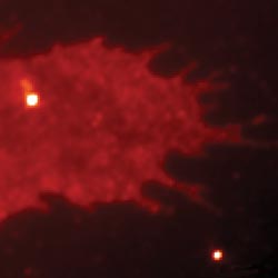
Figure 1. Traditional fluorescence microscopy is relatively low in resolution, revealing only the general location of fluorescently tagged molecules within a cell. Images courtesy of Harald Hess.
With PALM, however, the labels are not all energized at once, but in smaller sets. As each set is irradiated by the external light source, an image is acquired. For each cell of interest, thousands of images are acquired, each with a different set of fluorescent tags set to glow. The result is a single compiled image with a resolution as low as 10 μm (Figure 2).
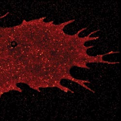
Figure 2. With photoactivated localization microscopy (PALM), developed at HHMI’s Janelia Farm Research Campus, fluorescent tags are rapidly switched on and off while thousands of images are acquired. The images then are combined into a single high-resolution picture.
PALM proved to be a great technique for deriving two-dimensional images of cells and their components, but cells don’t work merely in two directions. Hess began to work with other collaborators from Janelia Farm as well as from the National Institutes of Health in Bethesda, Md., and from Florida State University, Tallahassee, on using PALM to create 3-D images of cells.
He and his colleagues focused on bringing out the extra dimension by incorporating PALM with interferometric techniques. Interferometry typically uses data garnered from the paths of two light beams – usually a single laser beam passed through a beamsplitter – to measure how far an object has moved, the difference in height between adjacent objects or other spatial information.
Unfortunately, fluorescent particles inside a cell give off their own light, and you can’t readily bring a laser or other light source directly into the cell to provide the all-important reference beam. Hess and co-author Gleb Shtengel found the solution in quantum theory: The dual-wave-particle nature of a photon means that a single fluorescent molecule emits light in two optical paths simultaneously, providing its own reference beam.
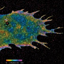
Figure 3. Bringing interferometry to PALM provides information on the location of tagged molecules throughout the cellular volume. The position of the fluorescent particles is color-coded, with purple molecules the highest, and red, the deepest.
Using this principle, the investigators set up the PALM system to collect light from the fluorescent labels both above and below the sample simultaneously. They used a three-way beamsplitter made by Rocky Mountain Instrument Co. to send the self-interfering emissions to separate cameras. The depth of each molecule in a cell determines how much light reaches each camera, which provides images such as in Figure 3.
The researchers show their work in a recent issue of the Proceedings of the National Academy of Sciences.