Unless a patient has worrisome risk factors or symptoms that are suggestive of heart disease, the American Heart Association (AHA) is advising against cardiac imaging tests that use radiation, according to its councils on Clinical Cardiology and Cardiovascular Radiology and Intervention. Risks from the ionizing radiation levels used in cardiac CT and nuclear medicine scans, although low, are not fully understood, and any unnecessary or repeated exposure could be damaging in the long run.
These findings by an AHA advisory committee led by Dr. Thomas Gerber were published in the Feb. 3, 2009, issue of the journal Circulation to make physicians and patients aware of possible health-related hazards.
Effective dose
The effective dose is a parameter measuring radiation’s potential harm to a patient. Estimating radiation dose as well as determining its health risks is complex because variables such as age, gender, medical history and intrinsic risk for cancer must be considered. Gerber, a cardiologist at the Mayo Clinic in Jacksonville, Fla., said the measurement is not as precise as doctors and patients would like to believe.
 It’s “nice to think that you have a handle on things, but…there is a false sense of security,” Gerber said. “Effective dose should be reported as a range rather than a specific number.”
CT imaging has become especially well accepted as a method for detecting plaque as an early sign of coronary artery disease in patients who have no symptoms. However, it has not been proved that identifying plaque early in the disease process will lengthen a person’s life.
According to Gerber, if a 45-year-old patient has only one or two risk factors such as high blood pressure or high cholesterol but no symptoms of heart disease, he is not a good candidate for cardiac CT, even if his coronary arteries reveal signs of plaque. On the other hand, a patient who has chest pain and risk factors for coronary artery disease is a justifiable candidate for the procedure. He could have undiagnosed heart disease that could lead to a potentially fatal heart attack. Thus, the benefit outweighs the radiation risk.
Lowering the risk
If, after a thorough examination, it is determined that a patient requires cardiac CT imaging, doctors can use certain strategies to lower the effective radiation dose and the potential risk to vital organs. In another study co-authored by Gerber and published in the Feb. 4, 2009 issue of JAMA, researchers examined four procedures that physicians can use to reduce the dose-length product (DLP), the total radiation produced by a 64-slice CT scanner to create a coronary CT angiogram (CCTA). This technique is widely used because it can detect coronary artery disease without an invasive catheter angiography, for example.
One way to reduce radiation is automatic exposure control. The feature adjusts the radiation output of the x-ray tube according to a patient’s individual anatomy. However, not all scanners have this, and it is not very effective in CCTA.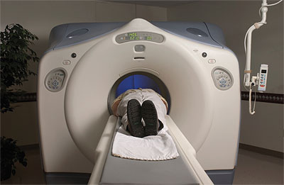
A second method is electrocardiographically controlled tube current modulation (ECTCM). During a predetermined time window of the diastolic phase of the cardiac cycle, when the heart is fairly still and images are less likely to be distorted, a normal tube current is maintained, whereas in the systolic phase, the tube current is decreased. However, if extra heartbeats occur, it’s possible that the scanner will apply a tube current that is incorrect for the particular phase of the cardiac cycle, so it’s important that the heart rhythm during the procedure be regular. In Gerber’s study, ECTCM reduced the DLP by 25 percent.
For CCTAs, the tube voltage usually is set at 120 kV, but researchers say that 100 kV also works well. This lower voltage is typical for pediatric CT scanning and for adults who are not obese; with larger patients, higher-dose radiation is needed to avoid image noise that may hinder a doctor’s ability to clearly read an image. In the study, tube voltage reduction, where possible, reduced the DLP by 46 percent.
Sequential scanning is another technique to lower radiation dose.
“The [method] that clearly reduces radiation the most is sequential scanning,” Gerber said.
This is when radiation is administered only at a particular time during the cardiac cycle. Throughout the rest of the cycle, the radiation tube is turned off, allowing for considerable reduction of radiation exposure. The study revealed a 78 percent decrease in the DLP. However, for this to work properly, the patient’s heart rate must be regular.
It has been suggested that, as imaging techniques advance and yield higher-resolution scans, collective and individual radiation doses will increase also. The advisory quoted from a recent report from the National Council for Radiation Protection and Measurements that, from 1982 to 2006, the overall collective radiation dose has increased significantly, by approximately 700 percent. In 2006, CT alone accounted for about 50 percent of the collective radiation dose, while nuclear medicine studies, which also are commonly used for cardiac imaging, have reportedly increased 5 percent annually. The 20 million nuclear medicine studies in 2006 represent about 25 percent of the overall collective radiation dose.
Therefore, it is important for physicians to exercise their best judgment about using such imaging tests and to weigh the benefits against the risks. Although there may be no argument that CT imaging is an important tool for studying cardiac symptoms, Gerber and the committees recommend that radiation exposure be avoided unless absolutely necessary and that it be used with proper radiation-lowering strategies appropriate for each patient.
Amanda D. Francoeur – News Editor
[email protected]