As the surgeon carefully prepares to make his first incision, he visualizes the route he planned and rehearsed earlier, guided by an ever-advancing suite of optical imaging tools. Thanks to optical molecular imaging, MRI, CT and the emerging field of optical coherence tomography (OCT), medical professionals are not only better equipped, but they also are better able to diagnose and plan procedures ranging from biopsies to surgery.
MRI and CT are commonly used in radiology to provide real-time subsurface images of patients noninvasively. Unlike CT, MRI uses non-ionizing radiation and provides much greater contrast between the various soft tissues of the body. But the relatively new world of OCT is where much of the cutting-edge research can be found.
OCT enables micron-scale, cross-sectional and 3-D imaging of biological tissues in situ and in real time. To date, OCT has broken new ground in ophthalmology, where it provides cross-sectional images of retinal pathology with higher resolution than any other noninvasive imaging technique.
Looking ahead, OCT is expected to make rapid progress in cancer detection, cardiology and dermatology, and in guiding surgical procedures. The technique is akin to ultrasound, except that imaging is performed by measuring light instead of sound. While imaging depths are shallow (around 1 or 2 mm) compared with those of ultrasound, the resulting resolution is considerably better (on the order of a few microns).
According to a 2010 market report on OCT by optics business analyst Strategies Unlimited, systems sales grew in 2009, despite the economic crisis, with a compound annual growth rate of 20 percent expected through 2014. The report also finds that Carl Zeiss Meditec of Jena, Germany, no longer holds the majority of the market share, even as its own revenue has increased. At least 21 other companies are developing and/or marketing OCT systems in eight medical specialties, as well as in R&D and industrial applications.
A giant in another field of 3-D imaging is Caliper Life Sciences of Hopkinton, Mass., which specializes in optical molecular imaging – a powerful tool for studying specific biomolecules and their real-time interactions in vivo. Strategies Unlimited reports that growth of equipment sales related to optical molecular imaging is on track to reach $400 million in 2014 and nearly $1 billion by the end of the decade.
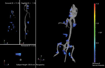
This 3-D image shows bioluminescent leukemia cells tracking to the lymph nodes in a mouse lymphoma model. Data courtesy of Caliper Life Sciences.
Unlike OCT, optical molecular imaging involves transgenic expression of bioluminescent proteins or tagging specific molecules or tissues with a fluorescent marker, which is then imaged via laser excitation.
“Caliper’s optical imaging products are used in basic research and in drug development in all of the major therapeutic areas, including oncology, infectious disease and cardiovascular disease,” said Dr. Stephen Oldfield, who is senior director of marketing for imaging products at Caliper Life Sciences. “For example, Caliper’s IVIS Spectrum is used for high-throughput 2-D imaging or 3-D imaging of small animals using either fluorescence or bioluminescence or both.”
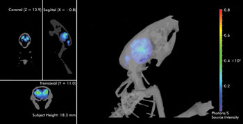
Shown is a 3-D image of bioluminescence due to GFAP protein expression in the mouse brain, induced by an inflammation response to prion infection. Data courtesy of Caliper Life Sciences and the University of California, San Francisco.
According to Oldfield, the system visualizes fluorescent or bioluminescent proteins, dyes and reactive probes using the most sensitive camera available, and IVIS systems are all calibrated to NIST standards so that data can be quantified and compared over a longitudinal study and between experiments.
Delving into 3-D fluorescence imaging of deep tissues requires transillumination, where excitation light is supplied to one side of the animal, and the emission light is collected at the other. This is then followed by a full tomographic analysis of the data.
This 3-D image shows bioluminescent leukemia cells tracking to the lymph nodes in a mouse lymphoma model. Data courtesy of Caliper Life Sciences.
“Just viewing the surface from different sides cannot tell you about the location of a signal deep within the animal,” Oldfield said. “When multiple optical signals such as bioluminescence and several fluorescent reporters are used together, they can shed light on some complex biological problems.”
At first, IVIS Spectrum could report on the growth of a tumor or the spread of an infectious disease. Now, however, the system can interrogate the underlying biology and follow multiple events during those processes. Oldfield predicts that in the next five to 10 years, new probes will extend the field even further.
“Tissue-specific transformation will enable biologists to investigate more complex signaling pathways and subtle modulation of gene expression in whole animals,” he said. “New reactive dyes will report on multiple enzyme activities using changes in their fluorescent properties. All of these will co-register with other imaging modalities and, ultimately, probes will be available for use in the clinic.”
Software: 3-D’s unsung hero
Of course, software is critical to making these developments possible but is often overlooked and underrated. It is the software that makes sense of the overwhelming amount of image data, presenting it clearly and quickly.
“A CT or MRI scan can consist of one thousand or more axial slices, and navigating through these slices can be simplified by having a 3-D view,” said Henri Primo, director of marketing and strategic relationships, Image and Knowledge Management at Siemens Healthcare. “Three-D visualization is a key technology for understanding these large data sets and how the anatomy under examination relates to other surrounding anatomy in 3-D space.”
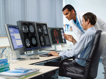
Radiologists review a 3-D reconstruction of the carotid arteries using Siemens syngo imaging IT software. Image courtesy of Siemens Healthcare.
Although the radiology community has been familiar with 3-D imaging for many years, employing the technique in daily operations remained a challenge for some time. Before it could become a staple tool, dedicated and expensive 3-D workstations had to be deployed, images had to be routed from the CT or MRI workstation, and data sheets had to be cleaned up.
Multislice/multidetector CT examinations continue to advance, with multislice capabilities increasing from four to 256 slices per scan. Study size has also increased from 50 to 500 MB, or even to more than 1 GB. Simultaneously, acquisition time for one exam has decreased from minutes to seconds.
“CTs can now be used not only for imaging static anatomy such as the brain or spine, but also to acquire dynamic studies of the heart,” Primo said. “We can now scan large anatomical areas in volumetric, isotropic modes, an application often used in cardiac scanning.”
What’s more, radiologists can now automatically format 3-D data sets according to their preference, depending on the type of user specialty and targeted disease. Siemens optical systems also offer continual software updates and live access to the data from any location.
While 3-D and advanced visualization tools are already being used for surgical planning and surgical guidance, the application of 3-D in medical imaging is expected to grow into other fields, such as pathology and cardiology.
The deployment of these technologies is also becoming more flexible and secure thanks to thin-client technology, which promises to eliminate the need for dedicated 3-D workstations. The notion involves a central server that provides preprocessing and floating license management.
“The floating license concept allows concurrent users to access the system simultaneously, supporting their processing, viewing, reading and information-sharing needs,” Primo explained. “For the physician, this means information can be viewed using thin-client devices for 3-D display, such as via a handheld computer or cell phone.”
Meanwhile, Caliper’s latest software, Living Image 4.0, is also helping make 3-D imaging easier for routine analysis. According to Oldfield, 3-D imaging was once in the realm of programmers and “imaging jocks” to generate that one picture for publication. But now, using simplified menus and wizards in Living Image, it is easy for biologists to process their own data and generate compelling 3-D images in routine analysis.