
Diagnosis through light, not the needle
Radiologists one day
could have an IR-based diagnostic tool integrated into traditional ultrasound systems
that could lower the rate at which women must undergo breast biopsies for suspicious
lesions.
The noninvasive alternative to conventional needle biopsies is
based on an emerging imaging technique – diffuse optical tomography –
combined with ultrasound localization. In a study involving 178 women, Dr. Quing
Zhu, a professor of bioengineering at the University of Connecticut, and her collaborators
found that the two techniques can be used as an adjunct to diagnostic ultrasound
for distinguishing early-stage invasive breast cancers from benign lesions.
Today, when mammography and ultrasound cannot determine whether
a suspicious breast lesion is malignant or benign, physicians typically recommend
a needle biopsy to extract samples of the suspicious tissue for laboratory testing.
In current clinical practice, the majority of biopsies performed reveal benign lesions,
leading to unnecessary anxiety for women.
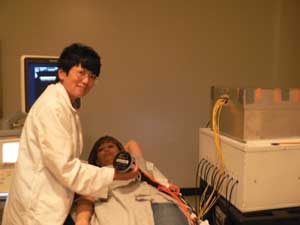
Dr. Quing Zhu is shown with a patient in a radiology exam room at the University of
Connecticut. The commercial ultrasound machine can be seen on the left. On the right
is the near-IR imager designed and built by Zhu’s team. Courtesy of Dr. Zhu.
“The large number of biopsies performed for benign breast
abnormalities has long been recognized as a serious problem,” Zhu said. “Optical
tomography offers complementary functional parameters, such as tumor angiogenesis
[proliferation of blood vessels] and hypoxia [lack of oxygen], when compared to
conventional x-ray and ultrasound imaging techniques, and therefore has significant
potential to assist the characterization of benign and malignant processes and reduce
unnecessary, normal biopsies.”
Dead giveaway
A rapidly growing tumor gives itself away: It has many more blood
vessels than normal tissue and rapidly outgrows its blood supply, leaving portions
of the tumor with regions where the oxygen concentration is significantly lower
than in healthy tissue.
Diffuse optical tomography can measure light absorption within
tissue to quantify blood content (hemoglobin level) and blood oxygen levels and,
hence, help distinguish malignant from benign lesions.
In Zhu’s study, published in Radiology online in June 2010
and in the print edition in August, the investigators used ultrasound to locate
the suspicious lesion, then performed diffuse optical tomography by shining IR light
into the area and measuring light absorption at two optical wavelengths.
They computed total hemoglobin levels from the light absorption
measured at optical wavelengths of 780 and 830 nm and correlated the measurements
with biopsy results. Laboratory examination of tissue samples revealed two in situ
carcinomas, 35 carcinomas measuring less than 2 cm, 24 carcinomas measuring more
than 2 cm and 114 benign lesions.
“Our next logical step is to work with ultrasound manufacturers
to incorporate the light imager into commercial ultrasound units for conducting
prospective clinical trials at multiple hospital sites,” Zhu said.
Published: September 2010