Compiled by BioPhotonics staff
Minute neural eye cell structures can now be viewed in tissue at high resolution,
and researchers say the technique could bring clarity to the study of circuit patterns
in all regions of the nervous system.
A combination of two microscopy methods allowed researchers at
the Max Planck Institute to discover that the synapses between retinal ganglion
cells and starburst amacrine cells are distributed asymmetrically. When viewed from
the ganglion cell, the starburst cell dendrites connected with it run opposite to
the preferred direction of motion. Their work appeared in the March 10, 2011, issue
of Nature (doi: 10.1038/nature09818).
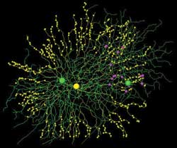
Cells and synapses reconstructed from serial block face electron
microscopy data. The image depicts a single starburst amacrine cell (yellow) and
two direction-selective ganglion cells (green). Although there is substantial dendritic
overlap with both cells, all connections (magenta) go to the correct ganglion cell.
Courtesy of Kevin Briggman, Max-Planck-Gesellschaft.
The scientists made the discovery by combining a new electron
microscopy method known as serial block face electron microscopy with two-photon
fluorescence microscopy. First, with the latter, they determined the preferred motion
direction of the ganglion cells. A calcium-sensitive fluorescent dye indicated the
stimulation of calcium flow into the cell – a process that signals electrical
activity within the cells.
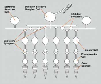
The optic signals in the outer segments of the photoreceptor cells are transduced into electrical signals. Excitatory and inhibitory synapses convey these signals to the ganglion
cell. Courtesy of www.somedonkey.com.
Then, using serial block face electron microscopy, they measured
the exact trajectory of all of the ganglion cell’s dendrites, as well as those
connected to amacrine cells. The new process used an electron beam to produce a
volumetric image by repeatedly scanning the surface of a tissue sample. Using an
extremely sharp diamond knife, they “sliced” the sample surface after
each scan.
Tracing the fine, densely packed branched dendrites of retinal
neurons, the scientists clearly identified their synapses. The complete automation
of the imaging process enabled them to record data sets with up to tens of thousands
of sections.