Multiphoton microscopy techniques –
including two-photon fluorescence and single-harmonic generation (SHG) – have
blossomed over the more than 20 years since they first became widespread, but they
continue to evolve, becoming more useful than ever to biologists. Much of the new
work being performed with multiphoton techniques is additive: using adaptive optics,
for example, or combining it with an adjunct methodology such as coherent anti-Stokes
Raman scattering (CARS)-based spectroscopy. The following are a few examples of
exciting work centered on these multimodal methods.
Sorting signals
Optical coherence microscopy (OCM), which offers high-lateral-resolution
structural information, and multiphoton microscopy (MPM), which provides high contrast
and submicron-resolution functional data, are a natural fit. The two techniques
highly complement each other, but the combined systems tend to be quite large, primarily
because it has been difficult to switch MPM to work with fiber lasers. Pulse broadening,
weak signals and nonlinearity have plagued such attempts.
Now, however, a team of researchers at the University of California,
Irvine, has developed a fiber-based Fourier-domain OCM/MPM system that operates
off a single laser source. The result is a compact multimodal system that could
lead to clinical use of endoscopic imaging at the bedside.
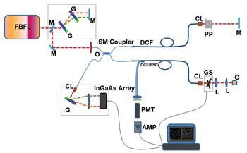
Figure 1. This schematic shows how researchers at the University of California, Irvine,
combined optical coherence microscopy (OCM) with multiphoton microscopy (MPM) techniques.
FBFL = fiber-based femtosecond laser; AMP = preamplifier; CL = collimator; G = grating;
GS = galvanometer mirror; M = mirror; O = objective; PMT = photomultiplier tube;
PP = prism pair; SM = single mode. Courtesy of Gangjun Liu.
Led by Gangjun Liu, the Irvine team based its system on a lab-built
fiber-based femtosecond laser emitting 80-fs pulses at a wavelength of 1.04 µm and
a repetition rate of 76 MHz (Figure 1). Crucial to the ability to adopt a fiber
laser, however, is that the system incorporates a (2+1):1 pump/signal combiner.
The device features a pair of multimode fibers fused to a double-clad fiber, which
effectively permits the proper light paths for the signals necessary for both OCM
and MPM operation (Figure 2). Double-clad fibers typically are used in multiphoton
applications because of their collection efficiency.
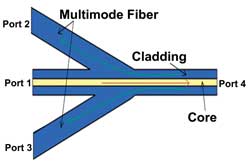
Figure 2. This schematic illustrates the light paths through a (2+1):1 pump/signal combiner, which makes it possible for OCM and MPM techniques to use the same light source.
Reused with permission of the Journal of Biomedical Optics.
The collection efficiency for the MPM signal is 20 percent using
this combiner, but the group reports that other combiners – with configurations
up to (19+1):1 – could collect as much as half the MPM signal. The OCM and
MPM paths do not at all affect one another, despite the use of a single-fiber light
source.
Liu and his colleagues successfully used the system to obtain
OCM, MPM and second-harmonic generation images from a variety of samples, with the
multimodal system providing both structural and functional information. They now
are working to further reduce the size of the probe to less than 2 mm and are testing
fiber lasers of various wavelengths.
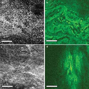
Figure 3. Shown are OCM images (a, c) of fixed and stained rabbit
heart tissue along with single-harmonic generation (SHG) images (b, d) of a dense
rat tail tendon. Image reused with permission of the Journal of Biomedical Optics.
In your eye
Two-photon excitation fluorescence is an excellent method for
studying tissues, especially in the eye, where there is higher transparency. The
technique provides the ability to see samples in sections along the Z-axis, offering
insight into both healthy and diseased components at various levels within the depths
of a tissue section.
“Very different cells and structures appear at different
depth positions,” said Juan M. Bueno of Universidad de Murcia in Spain. “It
is especially interesting for basic research to analyze the ocular structures under
intact conditions and visualize them with optimized contrast and resolution.”
The technique, however, is particularly affected by the intrinsic
wavefront aberrations in the microscope’s optics. Aberrations such as defocus
and astigmatism are the most common causes of poor image quality, although defocus
does not play a role in microscopy. Researchers at various institutions have tried
to correct astigmatism and other aberrations using software to alter the shape of
a deformable mirror or liquid crystal modulator in the optical path and have found
some success with fixed and stained samples ex vivo. Until recently, however, none
used a wavefront sensor to close the loop between aberrations in the emission wavelength
and the corrective mirror or modulator.
To bring two-photon fluorescence techniques to the realm of in
vivo ocular research, Bueno and his colleagues needed to obtain the best performance
from the corrective tool – in their case, a 140-element deformable mirror
coated with gold for protection from the high-power laser they used.
Other researchers have focused on correcting aberrations generated
within samples without knowing the amount of aberration at each plane they looked
at, Bueno said. Instead, they used basic algorithms to change the shape of the mirror
or modulator in their systems.
“In our work, we measured and compensated for the exact
aberrations of the laser beam,” he said. The rest of the optics and the tissue
itself cause wavelength aberrations, but the group focused on the illuminating beam.
“Although this is only a portion of the total aberration of the entire system,
we were able to significantly improve the quality of the images (of ocular tissues).”
Bueno and his collaborators, Emilio J. Gualda and Pablo Artal,
based their imaging system on a Nikon TE-2000U inverted microscope, a 760-nm Ti:sapphire
laser, a Hartmann-Shack wavefront sensor made by Thorlabs Inc. of Newton, N.J.,
and a deformable mirror from Boston Micromachines Corp. of Cambridge, Mass. (Figure
4).
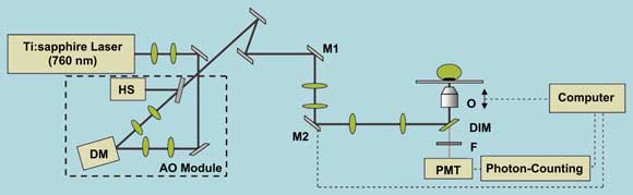
Figure 4. Introducing a Hartmann-Shack wavefront sensor (HS) and
deformable mirror (DM) into a multiphoton microscope system enables sharper imaging
of ocular tissues. AO = adaptive optics; M1/M2 = galvanometric mirrors; PMT = photomultiplier
tube.
As described in the November/December 2010 issue of the Journal
of Biomedical Optics, the researchers tested their system on intact human and porcine
eyes as well as on fixed and nonfixed ocular tissues. They found that the corrected
beam provided a better point spread function for image generation and permitted
the use of lower laser power, which will be essential for future in vivo ocular
studies in patients (Figure 5).
“In a few years … adaptive optics multiphoton microscopy
might be used in living eyes, if not for retinal imaging, at least for corneal tissue
analysis,” Bueno said.
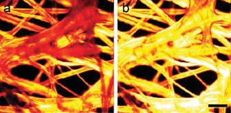
Figure 5. Two-photon excitation fluorescence images of a sample of fluorescent paper are shown before (a) and after (b) adaptive optics equipment corrected wavefront aberrations in the laser beam. Scale bar = 70 μm. Reused with permission of Journal of Biomedical Optics.
Next, the investigators intend to increase the depth of focus
of the system by manipulating the beam aberrations. They expect that doing so will
reduce the signal at the best-imaged plane but will provide higher contrast at other
levels along the Z-axis, Bueno said.
Under your skin
Digging through layers is not just the concern of ophthalmologists.
Skin also is of great concern, and research into better ways to examine skin layers
in situ – no tagging, no biopsies – is supported by the pharmaceutical
and cosmetic industries alike.
MPM, and the closely related multiphoton tomography, are useful
for imaging everything from single cells to whole animals, said Karsten König,
the head of JenLab GmbH in Jena, Germany, and a researcher affiliated with both
the University of California, Irvine, and Saarland University in Saarbrücken,
Germany. Neither technique, however, can find nonfluorescing biomolecules such as
lipids, water and hemoglobin. To get that information, König and his colleagues
add CARSto the mix.
“The combination of fluorescence/SHG (for collagen)/CARS
is the most interesting approach to image the most important biomolecules as well
as exogenous pharmaceutical substances and cosmetics,” he said.
Multiphoton/CARS tomography provides information on the diffusion
of materials through the stratum corneum (the outermost layer of skin), in situ
pharmacokinetics and the stimulated synthesis of collagen, among other interactions
between drugs and cosmetics with skin at contact and beyond, König said.
In an article published March 1, 2011, in the online edition of
Laser Physics Letters, König and his colleagues at JenLab and at the Berlin-based
APE GmbH and Charité (the medical school for both Humboldt University and the
Free University of Berlin) described the first use of a clinical CARS/MPM/SHG skin
biopsy system on human patients.
The group used a commercial multiphoton tomography instrument
made by JenLab. The overall system is based on a near-IR femtosecond laser operating
at 80 MHz combined with an optical parametric oscillator (OPO) emitting in the 1000-
to 1300-nm range. CARS stimulation came from femtosecond pulses from both the laser
and the OPO. The researchers kept the power below 50 mW to avoid tissue damage.
They examined the skin of healthy volunteers, patients with psoriasis and people
who had omega-3 oil applied to their otherwise healthy skin.
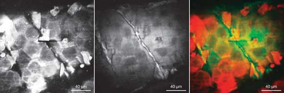
Figure 6. Researchers recently reported the first CARS/MPM imaging performed on human volunteers. Here, two-photon fluorescence imaging (left), CARS (center) and an overlay of the
two techniques (right) are shown. Scale bar = 40 μm. Reused with permission
of Laser Physics Letters.
Overall, the researchers reported that the resulting images provided
detailed information on several skin factors, including tissue structure and cell
morphology (Figure 6). They obtained good discrimination in psoriasis patients between
nonfluorescent lipids and water, and fluorescent molecules such as coenzyme NAD(P)H,
melanin, elastin and keratin.
JenLab already has sold several of its multiphoton tomography
systems but is working to enhance the system to detect more chemicals in the skin
as well as to further shrink it for clinical use.