Lasers are an established surgical tool in an ophthalmologist’s armory against glaucoma, cataracts and macular degeneration, but developments in OCT will have doctors turning to lasers before the patient ever reaches the operating room.
The development of lasik surgery has led to a rapid growth in the
vision correction market over the past two decades. But lasers are now starting
to find a place in the early detection and diagnosis of certain eye conditions,
particularly when used in optical coherence tomography systems.
The first application of femtosecond lasers in ophthalmology was
for lasik refractive surgery procedures, in which incredibly short pulses (around
700 fs) give surgeons the accuracy and predictability necessary for delicate tissue
vaporization.
During a procedure, a femtosecond laser is used to create the
corneal flap, approximately 150 µm thick, which is lifted prior to application of
an excimer laser that remodels the shape of the cornea.
According to Graham Cox, a professional services manager at Ultralase,
one of the leading refractive surgery providers in the UK and Ireland, the femtosecond
laser creates corneal flaps, which are thinner and planar (of even thickness) compared
with manual incisions. What’s more, the complication rate is greatly reduced,
from around 1 in 300 using a traditional bladed microkeratome, to 1 in 2500 using
a laser.
There are six companies that manufacture and sell femtosecond
lasers for ophthalmology: Abbott Medical Optics of Santa Ana, Calif., which acquired
Intra-Lase in 2008; Ziemer Ophthalmic of Port, Switzerland, which produces the FemtoLDV
femtosecond laser; Carl Zeiss Meditec of Jena, Germany, which offers the VisuMax
laser; Technolas Perfect Vision of Munich, Germany, which offers the Technolas Femtosecond
Workstation; Alcon Laboratories of Fort Worth, Texas, which acquired LenSx Lasers
in 2010; and LensAR of Winter Park, Fla., which sells the LensAR Laser System.
“Of these, the Intralase laser currently holds the overwhelming
market share, although this may change in the future as the other lasers are developed
for additional specialist applications,” Cox said. “Although there have
been minor enhancements to this [Intralase-based lasik] application, such as increased
frequency of the laser, giving a consequent reduction in the treatment time, it
is likely that a plateau has been reached in this field, given the very high accuracy
and predictability of modern lasik procedures.”
In 2010, Carl Zeiss Meditec introduced a new approach to refractive
surgery using its VisuMax laser to correct myopia and myopic astigmatism. Rather
than ablating corneal tissue with thousands of excimer laser pulses, the femtosecond
lenticule extraction (ReLEx) procedure enables tissue to be removed in one piece
through either a complete flap opening or a small 3- to 5-mm incision.

The first step in the ReLEx procedure is to anesthetize the eye using special drops. The VisuMax femtosecond laser then creates a thin lenticule in the intact cornea
and makes an access cut for its removal. Once the surgeon manually removes the lenticule
to change the refractive power of the cornea, the top corneal layer is flipped back
into its original position. Images courtesy of Carl Zeiss Meditec.
“The disadvantage of lasik treatments is that the cornea
has to be fully opened by the flap. This may potentially result in biomechanical
instability. In addition, the opening leads to the dry eye syndrome in some individual
cases,” said Dirk Mühlhoff, head of refractive lasers at Carl Zeiss Meditec.
“For this reason, we at Carl Zeiss Meditec sought a minimally invasive approach
using the femtosecond laser to cut the tissue to be removed from the cornea in a
single piece called an intrastromal lenticule. There is no longer any need to create
a flap, and the cornea is protected.”
The new approach is already marketed, and Mühlhoff reveals
that extraction of the corneal lenticule through a tiny keyhole incision is scheduled
for market launch in the second half of 2011.
Diagnosis and surgical guidance
The growth in OCT imaging procedures over the past 10 years has
been astounding, said Eric Swanson, one of the co-inventors of OCT and co-founder
of Advanced Ophthalmic Devices (an ophthalmic OCT imaging company that was acquired
by Carl Zeiss Meditec and formed the basis of Zeiss’s ophthalmic OCT product
line) as well as editor of www.octnews.org, an OCT news site.
Swanson and Dr. David Huang, OCT co-inventor and a leading researcher
in the field, calculated that for US Medicare patients, there were fewer than half
a million procedures related to OCT in 2000, but by 2010 this figure had reached
nearly 9 million. Worldwide, they speculate that more than 20 million clinical OCT
scans were carried out in 2010 and billed at a cost of more than $1 billion.
The meteoric rise can be attributed in part to the fact that OCT
enables cross-sectional and volumetric imaging of the retina and cornea at resolutions
that cannot be achieved by any other means except biopsy and histology. The images
that OCT provides dramatically improve the ability to identify retinal diseases
at an early, treatable stage.
In addition to being accepted as a standard diagnostic technique
by ophthalmologists, it is also rapidly gaining acceptance by optometrists.
“Today there are over 10 companies supplying OCT systems
for the ophthalmology and optometry markets. These companies are in fierce fights
for market share and are investing lots of development dollars to achieve differentiation
and drive sales,” Swanson said.
“As a result, there have been numerous advances in speed,
accuracy, higher lateral resolution, higher longitudinal resolution, image processing,
compactness, ease of use, functional imaging (e.g., Doppler detection) and lowering
capital equipment costs. This has brought, and will continue to bring, benefits
to both physicians and end patients,” he continued.
For OCT pioneer and professor James Fujimoto at MIT, one of the
most exciting developments in the technology is the use of narrowband swept lasers.
Swept lasers enable operation at long wavelengths such as 1000 and 1300 nm, where
high-speed camera technology is not readily available.
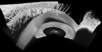
Ultrahigh-speed OCT imaging enables 3-D volumetric data sets to
be acquired. Images of the anterior eye (shown here) enable measurement of refractive
parameters of the eye. Courtesy of MIT.
“These long wavelengths have improved image penetration
in scattering tissues,” he said. “In ophthalmology, there is research
interest in using swept-source OCT at 1000 nm because it is less sensitive to cataracts
and enables imaging in scattering tissues, such as the choroid, the vascular network
behind the retina.”
Swept-source OCT can image around 10 times faster than commercial
spectral OCT instruments, or almost 1000 times faster than the first-generation
OCT technology, which used superluminescent diode light sources.
Since swept-source OCT requires the use of a narrow-bandwidth
light source that can be frequency swept at high repetition rates, there has been
a renewed research interest in tunable light sources.
“There are new developments in swept-source OCT using MEMS
[microelectromechanical systems] tunable vertical-cavity surface-emitting lasers
(VCSELs),” Fujimoto said. “In many ways, the VCSEL is an ideal light
source for OCT. It operates with a single longitudinal mode and can be rapidly tuned
using a MEMS mirror. The linewidth is very narrow, so it is possible to perform
OCT imaging over centimeter-depth ranges.”
What’s more, because tuning speed is limited in part by
the cavity lengths, the short cavity of the VCSEL enables it to tune at record speeds.
Praevium Research in Santa Barbara, Calif., together with Thorlabs
in New Jersey and Fujimoto’s group at MIT, recently demonstrated OCT imaging
with 760-kHz axial scan speeds as well as record imaging depths. The group presented
its work at CLEO 2011. According to Fujimoto, the VCSEL can be scaled to operate
in different wavelength regions and is a promising light source for future-generation
OCT.