The use of IR and UV-VIS spectroscopy enables safer, noninvasive analysis of soft tissues, and can enhance accuracy and speed in clinical diagnostics and medical research.
Spectroscopy is leading the way in both health care and medical research as a novel tool for improving diagnostics, disease detection and sensing. Dozens of spectroscopic techniques have evolved since the Indian researcher Sir C.V. Raman discovered a spectroscopic technique to observe molecular vibration and rotation in the 1920s. In more recent years, several exciting advances have made spectroscopy across the spectrum from near-IR to UV-VIS particularly invaluable for its promise in biomedical applications.
Near-IR spectroscopy (NIRS), from approximately 800 to 2500 nm, is an emerging imaging technology with “potential to redefine medical diagnostics and clinical research,” according to a 2013 market research report by Research and Markets.1 Scientists have used NIRS data from photon absorption and scattering for decades to identify different types of chemicals and compounds. In more recent years, use of the technology has advanced to biomedical applications for its advantages over existing radiological techniques; spectroscopy at IR and visible wavelengths avoids the use of ionizing radiation, and it is noninvasive. Although NIRS is in an early stage of commercial development, it may enable enhanced accuracy in clinical research and pathological diagnosis in the next five years, the report says.
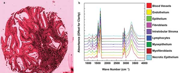 Mid-IR spectroscopy and interactive data-mining techniques (a) enable faster identification of the CH functional groups in colon tissue (b). Courtesy of R. Bhargava, University of Illinois at Urbana-Champaign.
The data angle
A new wave of data manipulation and interpretation in IR imaging will enable leaps in biomedical research and diagnosis, according to Rohit Bhargava, professor of bioengineering at the University of Illinois at Urbana-Champaign and winner of the 2013 Craver Award for pioneering IR spectroscopic imaging.1,2 Bhargava and his team developed a method to improve identification of mid-IR spectral features in cancer cells using advanced data manipulation. In a recent paper, the team describes a method of interactive data mining of spectral features obtained at mid-IR wavelengths using GPU (graphics processing unit)-based manipulation (doi: 10.1186/1471-2105-14-156). The proposed software enables a trained user to quickly separate, visualize and identify spectral features by tissue type. The tissue characteristics can then be used to obtain a much higher-resolution image, leading to faster tumor diagnosis than older methods allowed, without the use of a chemical stain.
“Spectroscopic imaging allows us to examine both morphology as well as molecular content in tissues, without perturbing the sample,” Bhargava said. “This presents a new opportunity for molecular pathology directly from the spectral data as well as synergistic integration with other assays. Exciting results from many labs have shown that this can improve diagnoses, eventually leading to better patient outcomes.”
Scientists at Boston University (BU)and the University of California, Irvine, have also focused on streamlining data: in particular, via the use of digital hardware. Dr. Darren Roblyer, assistant professor of biomedical engineering at BU, led colleagues in an effort to construct a relatively simple, faster and less-expensive all-digital spectroscopy system. The use of off-the-shelf digital technology instead of bulky benchtop equipment and complex analog circuitry enables improved wavelength-multiplexing schemes – and a much smaller system at a fraction of the cost. The technique, called frequency-domain diffuse optical spectroscopy (DOS), uses near-IR lasers modulated at radio frequencies (RF) to generate a photon density wave that traverses a sample of interest such as human tissue. The amplitude and phase of these waves are then detected and analyzed with high-speed all-digital sampling methods to determine the optical properties of the tissue, which can be used for medical diagnostic or prognostic purposes. Direct digitized sampling of raw data is made possible by recent advances in the speed of analog-to-digital conversion (ADC).
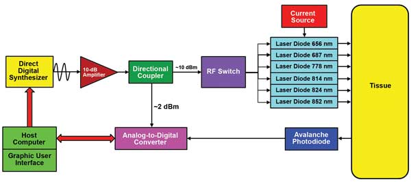 Direct digital sampling diffuse optical method: A direct digital synthesizer generates a broadband sweep of radio-frequency (RF) signals that sequentially modulate a series of near-IR laser diodes. An avalanche photodiode measures the frequency-domain optical signals after re-emission from tissue. An ultrafast analog-to-digital converter directly digitizes the signals, and Fourier analysis is used to calculate the phase and amplitude of measured signals, which reveal the type of tissue in the sample. Courtesy of D. Roblyer, Boston University.
“Our scheme has the potential to lead to faster, more portable, lower-power-consuming devices,” Roblyer said. “DOS may enable new medical and basic science applications, and may open new avenues for commercialization. The DOS technique can probe tissues more deeply – up to several centimeters – compared [with] other optical methods such as optical coherence tomography (OCT) and visible spectroscopy.” Potential applications include breast cancer diagnosis and chemotherapy monitoring.
“This technique provides a new design option for those seeking to make their own device – a much cheaper option than buying a $15,000 network analyzer or hiring an RF engineer to design a custom analog circuit board,” Roblyer added.
The team’s next goal is to greatly improve the measurement speed capabilities of the digital technique.
“By controlling the digital synthesis chips and ADC at the hardware level rather than at the software level, we hope to achieve very high measurement speeds that may allow us to explore biological phenomenon in a new way,” Roblyer said.
Better Raman
Raman spectroscopy is a powerful tool for tissue classification and disease diagnosis, but its clinical application faces considerable challenges. Hardware advances in components including lasers, filters and detectors have enabled scientists in St. Andrews, Scotland, and Jena, Germany, to demonstrate wavelength-modulated Raman spectroscopy as a viable method in real-time assessment of tissues in the operating room (doi: 10.3233/BSI-120031). To overcome the clinical issues of ambient light, background fluorescence and “etaloning,” a spectral distortion caused by the CCD sensor, Dr. Christoph Krafft at the Institute of Photonic Technology in Jena and colleagues recorded Raman excitation spectra of liver tissue at multiple slightly shifted wavelengths. The fact that Raman signals vary by wavelength, whereas the unwanted noise does not, enables the clean extraction of the Raman signature from the data. The hardware-based approach involves commercially available parts and requires a minimum of processing beforehand. With some improvements to sensitivity and throughput, the team expects the technique could soon enable practical in vivo tissue analysis in real time.
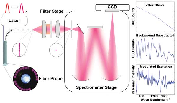 A prototype wavelength-modulated Raman spectroscopy system successfully suppresses unwanted spectral noise for real-time analysis of liver tissue during surgery. The setup includes a 785-nm modulated master-oscillator-power-amplifier (MOPA) laser, coupled to a commercially available fiber probe, spectrometer and CCD detector. Courtesy of Christoph Krafft, Institute of Photonic Technology.
Faster diagnosis is also the target of another study in Jena to identify urinary tract infection (UTI) pathogens using Raman microspectroscopy (doi: 10.1021/ac401806f). Professor Jürgen Popp, scientific director of the Institute of Photonic Technology, and colleagues have developed a proof-of-principle procedure to probe the spectroscopic fingerprint of single E. coli and E. faecalis bacteria, the most common UTI pathogens. The technique enables identification of the pathogens within approximately two hours of specimen collection, an enormous improvement over the standard 24-hour window for urine cultures, since it renders cultivation unnecessary. Further challenges remain. “To be a viable clinical option, we must enlarge the reference database to include more bacterial species, different cultivation conditions, and more strains for each bacterial species,” Popp said. “The parallelized and automated measurements of different samples must also be realized.” But these are surmountable goals that make the first single-cell typing of UTI pathogens possible in the very near future.
References
1. Research and Markets (2013). Optical imaging market (2013-2018) – forecasts to 2018.
2. R. Bhargava (2012). Infrared spectroscopic imaging: the next generation. Appl Spec 66, pp. 1091-1120. https://www.s-a-s.org/
journal/viewer/abstract/10810.
3. http://www.spectroscopyonline.com/podcasts
How spectroscopy works
Spectroscopy is used to study the structure of atoms and molecules by means of scattering and absorption of light into and from a medium. IR spectroscopy, from about 2500 to 16,000 nm in wavelength (or 1.9 × 10¹³ to 1.2 × 10¹⁴ Hz in frequency), causes vibrational excitation in virtually all organic compounds due to the unique component atoms and the covalent bonds in molecules. When light is directed through tissue or other soft organic material, the molecules within vibrate at characteristic frequencies reflecting their components.
The resulting spectrum works like a fingerprint to help scientists identify the components and study them. Spectrometry systems cover spectral ranges from near-IR to mid-IR and UV-VIS, depending on the chemical or molecule to be identified.
Trending now: Faster, better, smaller
With advances in off-the-shelf spectroscopy components and new data sampling and analysis techniques, research groups are making steady progress in improving tissue identification while reducing diagnosis times as well as spectroscopy system size and cost. The developed world can look forward to the benefits of these advances someday soon, and many researchers also hope that optical spectroscopy and imaging methods may ultimately fill unmet needs in developing nations, where in-field, portable, cost-effective systems could quickly scan for diseases like cervical or oral cancer.
 (a) Noninvasive UV-VIS spectroscopy can be used in real time to help assess the status and age of bruising in victims of possible physical abuse. (b) Optical spectroscopic sensing can help match ear prosthetics to natural skin tones. (c) Handheld Raman instruments such as the IDRaman mini from Ocean Optics are becoming smaller, more portable and more precise. Images courtesy of Ocean Optics.