Digital light microscopy has been demonstrated as a faster, easier method for conducting microindentation analysis — a tool often used to investigate the mechanical properties of materials such as bone.
Microindentation analysis of cross-sectioned bone samples can reveal differences in bone quality and fracture mechanisms. Since bone consists of a spongy structure, it is imperative to control the position of indentations using high-resolution imaging, ensuring that only the bone matrix was subject to indentation, rather than voids, pores or the polymer used for embedding the samples.
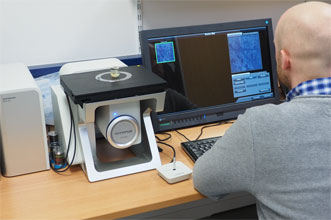
A team of researchers led by Dr. Björn Busse at the University Medical Center Hamburg-Eppendorf had been using scanning electron microscopy (EM) to investigate cross-sectioned bone samples embedded in polymer. To improve the efficiency of the microindentation process, Busse and his team performed high-resolution digital light microscopy as an alternative to EM, using the DSX500i inverted digital light microscope from Olympus Corp.
The DSX500i’s Best Image function allowed researchers to quickly find the best possible settings to reveal relevant details. Courtesy of E.A. Zimmerman and B. Busse, at al.
The microscope provided 13× zoom optics, and the combination of high resolution and operating simplicity improved the speed and efficiency of the microindentation process, the researchers said. Sample preparation time was especially reduced compared to that required with a standard light microscope.
When multiple indentations were made on each bone specimen, the DSX500i was quickly able to determine how many indentations hit bone, helping the researchers decide if more indentations were required.
Unlike electron microscopy, digital light microscopy can also provide depth information. For example, if the bone tissue was softer and had a lower mineral content or was harder and more brittle, that information was reflected in the depth of the indents.
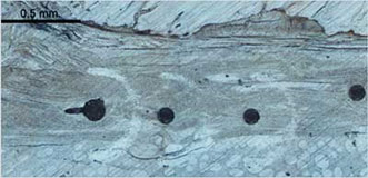
Four microindentations in cortical bone. This image was taken using the Stitching Function of the DSX500i. Courtesy of E.A. Zimmerman and B. Busse, et al.
Beyond indentation analysis, the researchers said the microscope presents other avenues of research, such as investigating the properties of cracks and their propagation that occurs adjacent to indentations. Additional applications of the technique could exist in other sectors of materials research.