Live-imaging of medaka fish — also known as Japanese rice fish — larvae aboard the International Space Station is helping scientists study the long-term effects of microgravity on the human body.
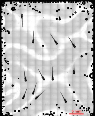
A total of 273
images captured by a 5× objective lens were integrated by the Tiling method, showing
an overall picture of the medaka chamber. This figure shows suitable localization
of medaka fish for observation by a 20× objective lens. Courtesy of Tokyo Tech.
Research shows that astronauts undergo a significant drop in bone mineral density during space missions, but the precise molecular mechanisms responsible for such changes in bone structure are unclear.
Using remote, real-time live-imaging, a team of scientists in Japan studied the fluorescent signals derived from osteoblasts — cells that make bone — and osteoclasts — cells that break down bone mass — of medaka fish after one day of microgravity exposure.
Using a modified fluorescence microscope, which was controlled on Earth, lead researcher Akiro Kudo and his team found increases in both osteoblast and osteoclast specific promoter-driven green fluorescent protein (GFP) and red fluorescent protein (DsRed) signals one day after launch; this continued for the next eight days. This signified that changes in bone mass happen almost immediately after launch in astronauts.
Kudo told Photonics Media being on Earth and remotely accessing the microscope on the International Space Station was challenging.
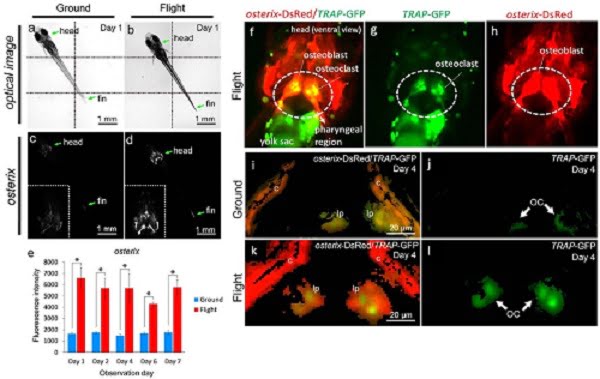
Bone
metabolism under microgravity — Increase of fluorescent signals of osteoblasts
and osteoclasts in medaka. (a-d)
Whole-body imaging of the osterix-DsRed
transgenic line. The left-side images show the same ground control at day 1; and
the right-side images, the same flight medaka at day 1. Arrows point to the
head and fin region. All images show ventral views. Montage images were made
from 6 captured optical images, divided by dotted lines (a,b). The white region
shows an osterix-DsRed
fluorescent signal. Embedded views show the enlarged head region (c,d). The
fluorescent intensity from day 1 to 7 of observation day constantly increased
in the flight group(e). The representative visualizing data for osterix-DsRed/TRAP-GFP in the flight
group (f-h). All images show ventral views in the head region. The merged
images were captured by 3D views for osterix-DsRed
and TRAP-GFP in
the pharyngeal bone region of the double transgenic line (i-l). The pharyngeal bone
region in the ground control (i) or the flight (k) group at day 4. The image
for TRAP-GFP in
the pharyngeal bone region of “i” (j) or “k” (l). lp, lower pharyngeal bone; c,
cleithrum. GFP signals identify osteoclasts (OC). Courtesy of Tokyo Tech.
“The microscope was controlled by specialized software that was able to
receive a command from ground control and then execute the microscope’s
actions continuously using a batch process,” said Kudo. “Fluorescent
signals were observed with the filter set and the LED light source
optimized each signal.”
The fluorescent analysis was complimented by using transcriptome — the set of all messenger RNA molecules in one cell or a population of cells — analysis to measure gene expression in the transgenic fish.

Medaka fish arrived at the ISS, and then were received by astronauts in 2014. Courtesy of Tokyo Tech.
Their findings suggest that exposure to microgravity induced an
immediate “dynamic alteration of gene expressions in osteoblasts and
osteoclasts. … [N]amely, these experiments based on real-time imaging of
medaka from Earth and transcriptome analysis could be the prelude to
the establishment of a new scientific area of research in “gravitational
biology, said Kudo.
During the next space experiment, Kudo and his colleagues will look at the role of glucocorticoid receptors (GR) on cells in microgravity, again using remote live-cell imaging techniques from the ground. The study results can be found in Scientific Reports (doi:10.1038/srep39545).