Innovations could enable real-time tissue analysis during prostate surgery, improved medication delivery for brain tumors and easy-to-administer tests for malaria.
Spectroscopy is no stranger to the life sciences. This noninvasive technique has been employed in the sector for decades. But now, researchers worldwide are finding new applications in the surgical suite and laboratories, as well as in disease detection and diagnosis.
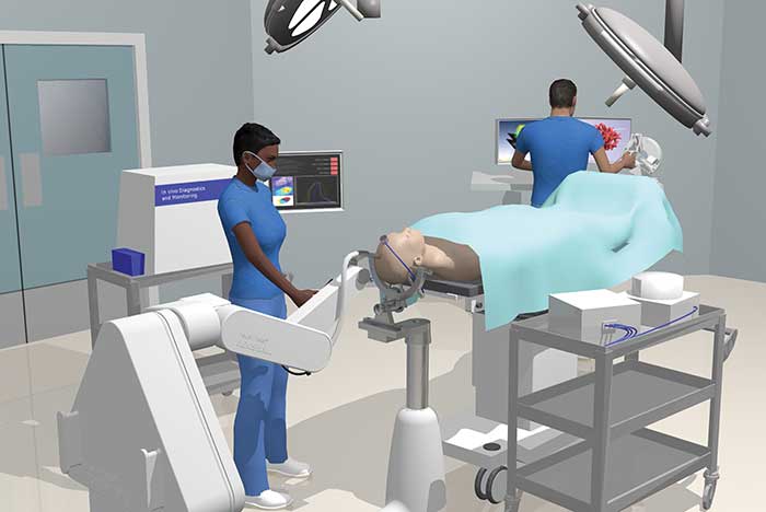
This illustration shows how a surgical procedure will look using the wasp-inspired robotic needle. Courtesy of Imperial College London.
Light-reflectance spectroscopy evaluates surgical margins
Light-reflectance spectroscopy is emerging as a formidable tool in the life sciences, and now it could enable real-time tissue analysis during prostate cancer surgery, as well as more accurate distinction between malignant and benign tissue.
In a study of patients with intermediate-to high-risk disease requiring radical prostatectomy, a team from the University of Texas Southwestern Medical Center employed a novel light-reflectance spectroscopy probe to evaluate surgical margins on radical prostatectomy tissue specimens and correlate the findings
with pathological examinations. Light-reflectance spectroscopy analysis was performed on 17 prostate gland specimens, of which a total of 11 histologically positive and 22 negative surgical margins were measured. The optical probe predicted positive surgical margins with 85 percent sensitivity, according to the researchers, in addition to 86 percent specificity and 86 percent accuracy.
Light-reflectance spectroscopy measures light intensity backscattered from tissues. In prostate cancer patients, a radical prostatectomy is often recommended. But due to the amount of time involved with traditional techniques and the lack of proven clinical usefulness, analysis to determine removal of all cancer surrounding the visible tumor’s edges is not routinely performed during surgery.
The benefits of the light-reflectance spectroscopy procedure, once fully qualified, could include highly accurate surgical removal of all cancerous tissue, the researchers said, as well as the ability to spare more healthy tissue, thus minimizing the likelihood of both cancer recurrence or additional treatment.
Further study is needed, however, according to UT Southwestern professor Dr. Jeffrey Cadeddu, “to determine whether such analysis may be used in real time to improve surgical decision-making and decrease the amount of tissue surgeons need to remove.”
FTIR spectroscopy at the subatomic level
Spectroscopy is making an impact in laboratory settings, as well. Specifically, Fourier Transform Infrared (FTIR), which, when combined with computer simulations, has been able to analyze and define at the subatomic level the mechanism that causes the proteins that control critical bodily processes to be switched off.
Using time-resolved FTIR spectroscopy, a team from Ruhr-University Bochum (RUB) was able to monitor enzymatic processes label-free at a high temporal and spatial resolution in their natural state and provide rate constants and structural information that were coded into IR spectra. In a study published in the Proceedings of the National Academy of Sciences (doi: 10.1073/pnas.1612394113), the researchers found that quantum-mechanical computer simulations of structural models aided in extracting information about the precise location in the enzyme where a process took place.
“By combining theory and experiment, we thus gain a microscope with subatomic resolution,” said RUB researcher Klaus Gerwert.
Bonded to the proteins, the energy molecule Guanosine-5’-triphosphate (GTP) is crucial for turning off many protein switches. If an enzyme cleaves a phosphate group from GTP, the protein switch is turned off. This so-called GTP hydrolysis takes place within seconds and is activated by a specific amino acid — the arginine finger. If the process fails, disease could develop. Using a combination of FTIR spectroscopy and bio-molecular simulations, the researchers were able to visualize the state of the arginine finger bonded to the GTP molecule at one hundredth of the atomic diameter, which presented in detail how GTP hydrolysis is accelerated.
“Our long-term aim is for our basic research to contribute to the development of drugs for the treatment of cancer and severe genetic diseases,” said researcher Daniel Mann.
Wasp-inspired needle for detecting disease
Raman spectroscopy is showing promise in assisting with the detection of disease.
Researchers at Imperial College London, in working with a robotic surgical system inspired by the anatomy of wood-boring wasps, have developed a prototype needle design based specifically on female parasitoid wasps, which uses a bendable needle-like ovipositor to bore into wood to lay eggs in hiding host larvae. The robotic needle could be used to analyze the body at the molecular level, using Raman spectroscopy to detect cancerous and diseased cells.
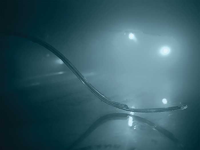
The wasp-inspired robotic needle is bendable, enabling it to access hard-to-reach areas deep in tissue. Courtesy of Imperial College London.
The technology consists of a minimum of three tiny interlocked polymer shafts that slide in parallel, mimicking the way the ovipositor works. The segments move in a complex pattern to minimize the needle’s impact on surrounding tissue, enabling it to travel along curved paths with dexterity and access hard-to-reach areas deep in tissue.
The new robotic needle will be mounted on a robotic arm, enabling it to be accurately positioned over a patient’s head. The system will intelligently guide the insertion of the needle alongside the surgeon, who researchers noted will control it using a haptic joystick — this will provide sensory cues to ensure the insertion is on target. Clinical trials are slated to begin at the end of 2020.
Imaging technique accurately diagnoses malaria
Quantitative phase spectroscopy shows significant clinical potential for detecting malaria and other infections without staining or expert analysis. A team from Duke University, in a study published in PLOS ONE (doi: 10.1371/journal.pone.0163045), used a quantitative phase spectroscopy (QPS) system to incorporate digital holography, which in turn has allowed identification of spot malaria-infected cells from a simple, untouched blood sample. And it has done so without any help from a human. The technique employs machine learning algorithms and has proved accurate in detecting malaria infection 97 to 100 percent of the time. The research could form the basis of a fast, reliable test for malaria that could be given by most anyone, anywhere in the field.
In the QPS-based system, the researchers said a laser sweeps through the visible spectrum, and sensors capture how each discrete light frequency interacts with a sample of red blood cells. The resulting data is captured in a holographic image that provides information indicating whether a malarial infection is present. The red blood cells were segmented via optical phase thresholds, while the samples were refocused to enable quantitative comparison of phase images. Refocused images were then analyzed to extract 23 morphological descriptors based on the phase information.
The researchers constructed machine learning algorithms, using the morphological descriptors of each cell taken from the quantitative phase images, in order to improve automated analysis of the red blood cells.
“We identified 23 parameters that are statistically significant for spotting malaria,” said Duke researcher Han Sang Park. “However, none of the parameters were reliable more than 90 percent of the time on their own, so we decided to use them all.”
Duke professor Adam Wax added that adoption relies on any new diagnostic device being “just as reliable as a trained field worker with a microscope.”
“Otherwise,” he said, “even with a 90 percent success rate, you’d still miss more than 20 million cases a year.”
The algorithms essentially combine all of the calculated physical parameters to distinguish cells more effectively and enable identification of malaria infection with a high level of accuracy, providing the ability to discriminate between stages of the infection.
“With this technique, the path is there to be able to process thousands of cells per minute,” Wax said. “That’s a huge improvement to the 40 minutes it currently takes a field technician to stain, prepare and read a slide to personally look for infection.”