Portable imaging and sensing devices can provide robust systems for both developing and developed countries.
QINGSHAN WEI, NORTH CAROLINA STATE UNIVERSITY, AND AYDOGAN OZCAN, UNIVERSITY OF CALIFORNIA, LOS ANGELES
Lack of diagnostic tools in resource-limited settings remains one of the major obstacles to disease diagnosis and treatment in the developing world. Although significant progress has been made in the availability of highly sensitive and specific molecular diagnostic tests, most of the current technologies are costly, relatively complex, and rely on advanced infrastructure and highly trained personnel, which restrict their applications to clinical laboratory settings. Even in developed countries, the cost of medical diagnostics can impose hurdles for patients.
Imaging and sensing devices based on smartphones have emerged to provide cost-effective, field-portable and robust diagnostic systems to meet global health and related biomedical challenges, both in developing and developed countries1,2. Furthermore, phone-based devices provide additional functionalities that are extremely useful for point-of-care (POC) applications. The phones’ self-contained computational power can enable real-time data analysis, spatio-temporal tagging and communication of results at the detection site. And various imaging and sensing attachments have been developed recently that can convert smartphones into advanced biomedical measurement tools.
Fluorescence microscopy
Many smartphone diagnostic applications have been achieved through fluorescence readout using a phone’s embedded camera. In general, the default camera lens of a smartphone is designed for capturing large-scale objects in ambient lighting conditions with a viewing angle of approximately 60°. An external lens can make the device suitable for imaging microscopic samples placed at a few millimeters to centimeters away. By forming a simple imaging system, the lens can project an image of a microscopic sample onto the active area of the CMOS imager chip installed on the phone.
Recently, researchers demonstrated that a good option for the choice of the external lens is a reversed smartphone camera lens3,4. This extremely cost-effective solution achieves both a decent spatial resolution and a large field of view (FOV).
Depending on the choice of external lens, low-cost smartphone microscopy tools can achieve sub-micron half-pitch resolution5 over a FOV of 3 to 4 mm2, and in some cases have a much larger FOV (~1 cm2)6,7, at the cost of resolution. This provides a highly versatile imaging tool that is well-suited for a variety of applications that include DNA imaging and sizing8, single virus detection3, high-throughput cell counting9, imaging flow-cytometry10,11, detection of parasites5 and pathology tissue slide imaging12.
For fluorescence imaging, an emission filter is needed. This can be placed in between the two lenses to reduce the angular dependency of the filter function, while also making the system compact. How the samples are illuminated is an important factor for enhancing the signal-to-noise ratio and, therefore, the detection sensitivity of the device. For volumetric samples introduced through cuvettes or tubes, an illumination path that is orthogonal to the detection path provides a simple means of significantly reducing excitation leakage13. For planar slide-based samples, waveguide coupling from the side of the glass slide also has been proven to be an efficient method6.
An alternative approach is based on oblique illumination of the sample slides to achieve better rejection of the excitation beam and improve the detection sensitivity (Figure 1a)3. In this configuration, the samples typically are back-illuminated by a laser diode beam. To suppress background leakage because of propagation and scattering of the excitation beam, the incidence angle of the excitation laser is selected to be significantly larger than the numerical aperture of the external lens. The design enables a microscope based on a smartphone to capture fluorescent signals from a few hundred fluorophores within a diffraction-limited spot. This is sufficient to detect a single fluorescently labeled virus particle (Figure 1b)3 or DNA strand (Figure 1c)8. The same design also works as a powerful dark-field microscope that can detect weakly scattering objects.

Figure 1. Smartphone fluorescence and dark-field microscopy based on an oblique illumination design can improve detection sensitivity. The schematic of the smartphone device shows a tilted laser illumination. Ray tracing simulation of the optical path of the imaging system is shown at the bottom (a). A smartphone fluorescence image shows isolated human cytomegaloviruses (green) (b). Another smartphone fluorescence image shows individual DNA strands (c).
Molecular pathology
One of the important applications of smartphone-based fluorescence microscopy is to read and quantify molecular assays and POC tests using cost-effective and mobile interfaces. Molecular pathology, where molecular analysis complements traditional morphology-based diagnosis of diseases, can greatly improve diagnostic accuracy and specificity, and provide valuable information for a wide range of medical conditions. It has recently been demonstrated that a multimodal smartphone fluorescence microscope can be used as a digital pathology tool for cancer point mutation analysis in tumor slides and on DNA array chips14.
Early detection and screening of cancer mutation is the key to more-efficient cancer therapy. However, the current gold standard diagnosis, which is based on polymerase chain reaction (PCR), is a complex process limited by a relatively slow turnaround time and lack of portability. A simple, cost-effective POC technology for genetic mutation detection is therefore an unmet need, especially for resource-poor settings.
The smartphone-based multimodal fluorescence microscope has successfully detected KRAS mutation in colon cancer patients14. The design features dual-color fluorescence detection (Cy3 & Cy5 channels) by using two compact laser diodes (532 and 638 nm) in the same attachment (Figure 2a). An additional white LED provides the third imaging channel in bright-field. The sample holder has been designed to enable the movement of the sample slide in three dimensions.
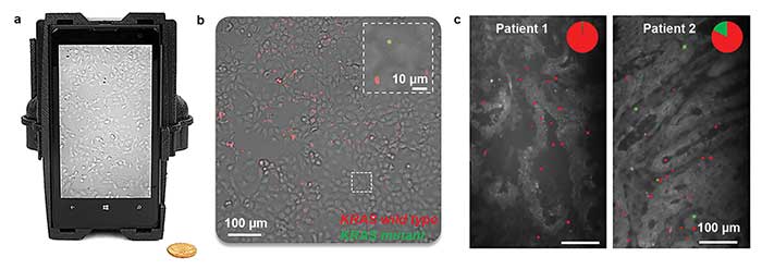
Figure 2. A multimodal smartphone fluorescence microscope can be used as a digital tool for cancer mutation detection (a). The smartphone image of in situ RCA assays in fixed model cell lines superimposes two fluorescent color channels onto the bright-field channel of the mobile microscope (b). The same multimodal smartphone microscope can image in situ KRAS mutation in colon cancer tumor sections (c).
Using this device, a KRAS diagnostic assay based on rolling circle amplification (RCA) and ligation chemistry were also developed to sequence a target DNA region containing the point mutation at the codon 12 position of the Ras gene. As a product of this RCA reaction, amplified single-stranded (ss) sequences form micron-sized DNA coils that can be labeled with distinctive fluorescent colors using short fluorophore-conjugated ss DNA probes. These, in turn, can be detected and counted digitally by using the smartphone microscope to quantify the ratio of the mutant to wild-type genes, each labeled with a different fluorescent color.
This concept was first tested by imaging synthetic or extracted DNA from model tumor cell lines on standard glass slides functionalized with capturing DNA ligands. The smartphone fluorescence microscope was able to detect as low as 1 fM target DNA sequence. A high sequencing depth (1:1000 mutant: wild type ratio) was demonstrated per FOV of the smartphone fluorescence microscope that is comparable to, if not better than, FDA-approved PCR-based KRAS diagnostic tests.
Furthermore, the same technology also was applied for in situ mutation detection in model cell lines or tumor tissue sections from patients (Figure 2b and c)14. The multimodal capability of the mobile microscopy platform allows spatial overlap of the mutation-related and wild-type fluorescence signals on top of the bright-field channel for multiplexed viewing. A specially developed machine learning algorithm achieves automated recognition of target signals against the nonspecific background. Overall, the smartphone device was able to accurately score all the patient tumor samples that were tested as KRAS positive or negative. The results were validated against the results obtained by next-generation sequencing of the same samples14.
Plasmonics-enhanced detection
Optical detection sensitivity is an important factor to further improve molecular diagnostic capabilities of microscopy and sensing devices based on smartphones. Better sensitivity might also reduce the assay time that is required. Toward this goal, a plasmonics-enhanced smartphone fluorescence microscope recently demonstrated ~10 times improvement in signal intensity using an easy-to-fabricate plasmonic substrate based on a thin silver film15.
The plasmonic imaging design uses a silver-coated (50-nm) glass slide as a substrate. Surface plasmon resonance (SPR) waves at the dielectric interface of the thin metal film enhances the local electromagnetic field and therefore the resulting fluorescence signal from target molecules. In this smartphone-based design, a compact Kretschmann illumination configuration was used by attaching a disposable silver-coated sample slide onto a glass hemisphere (Figure 3a). The p-polarized laser beam was then launched from the glass hemisphere side at an optimized incidence angle to provide decent coupling to plasmonic waves at the sample substrate. A silica-based spacer (~30 nm) balanced the fluorescence quenching versus signal enhancement. To improve the overall performance of the plasmonic system, a variety of parameters (metal film thickness, spacer distance, incidence angle, polarization, etc.) were optimized both experimentally and through numerical simulations, after which a smartphone-based prototype was created using 3D printing (Figure 3b).
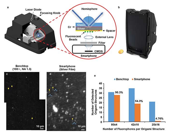
Figure 3. A plasmonics-enhanced smartphone fluorescence microscope makes use of an easy-to-fabricate silver-coated substrate shown in the schematics of the Kretschmann illumination configuration (a). The microscope fits neatly onto a smartphone (b). The individual 50-nm fluorescent particles detected by the device were validated by benchtop fluorescence microscope images, acquired using a 100× (NA 1.3) objective lens (c, d). The smartphone microscope’s detection limit was determined through the use of fluorophore-labeled DNA origami structures (e).
This optimized plasmonics-enhanced smartphone microscope allowed the visualization of single fluorescent nanoparticles as small as 50 nm in diameter (Figure 3c and d) as well as single quantum dots (~20-nm diameter) that cannot be detected using standard glass substrates. By using fluorescently labeled DNA origami nanostructures as brightness standards, the limit of detection of this mobile microscope was determined to be approximately 80 fluorophores per diffraction-limited spot (Figure 3e). The planar metal film generates a uniform field enhancement across the substrate area and also provides easy integration with microfluidic channels for more advanced sample preparation. This plasmonics-enhanced fluorescence mobile microscopy platform will be highly useful for a range of POC tests and molecular assays, bringing a highly sensitive and simple imaging and sensing tool to field and resource-limited settings.
Meet the authors
Qingshan Wei is an assistant professor at the Department of Chemical and Biomolecular Engineering, North Carolina State University; email: [email protected]. Aydogan Ozcan is a chancellor’s professor at the Electrical Engineering Department and Bioengineering Department, University of California, Los Angeles, and a professor at the Howard Hughes Medical Institute (HHMI); email: [email protected].
References
1. A. Ozcan (2014). Mobile phones democratize and cultivate next-generation imaging, diagnostics and measurement tools. Lab Chip, Vol. 14, pp. 3187-3194.
2. S.K. Vashist et al. (2014). Cellphone-based devices for bioanalytical sciences. Anal Bioanal Chem, Vol. 406, pp. 3263-3277.
3. Q. Wei et al. (2013). Fluorescent imaging of single nanoparticles and viruses on a smart phone. ACS Nano, Vol. 7, pp. 9147-9155.
4. N.A. Switz et al. (2014). Low-cost mobile phone microscopy with a reversed mobile phone camera lens. PLOS ONE, Vol. 9,
p. e95330.
5. I.I. Bogoch et al. (2017). Evaluation of a mobile phone–based microscope for screening of Schistosoma haematobium infection in rural Ghana. Am J Trop Med Hyg, doi: https://doi.org/10.4269/ajtmh.16-0912.
6. H. Zhu et al. (2011). Cost-effective and compact wide-field fluorescent imaging on a cell-phone. Lab Chip, Vol. 11, pp. 315-322.
7. H. Zhu et al. (2011). Optofluidic fluorescent imaging cytometry on a cell phone. Anal Chem, Vol. 83, pp. 6641-6647.
8. Q. Wei et al. (2014). Imaging and sizing of single DNA molecules on a mobile phone. ACS Nano, Vol. 8, pp. 12725-12733.
9. H. Zhu et al. (2013). Cost-effective and rapid blood analysis on a cell-phone. Lab Chip, Vol. 13, pp. 1282-1288.
10. H.C. Koydemir et al. (2015). Rapid imaging, detection and quantification of Giardia lamblia cysts using mobile-phone based fluorescent microscopy and machine learning. Lab Chip, Vol. 15, pp. 1284-1293.
11. H. Zhu et al. (2012). Quantum dot enabled detection of Escherichia coli using a cell-phone. Analyst, Vol. 137, pp. 2541-2544.
12. Y. Zhang et al. (2016). Color calibration and fusion of lens-free and mobile-phone microscopy images for high-resolution and accurate color reproduction. Sci Rep, Vol. 6, p. 27811.
13. A.F. Coskun et al. (2013). Albumin testing in urine using a smart-phone. Lab Chip, Vol. 13, pp. 4231-4238.
14. M. Kühnemund et al. (2017). Targeted DNA sequencing and in situ mutation analysis using mobile phone microscopy. Nat Commun, Vol. 8, p. 13913.
15. Q. Wei et al. (2017). Plasmonics enhanced smartphone fluorescence microscopy. Sci Rep, Vol. 7, p. 2124.