The unique spectral and temporal qualities of supercontinuum light sources are advancing our understanding of reactions in biology, chemistry and condensed matter.
ROBERT ALFANO, CITY COLLEGE OF NEW YORK, AND LINGYAN SHI, COLUMBIA UNIVERSITY
For many applications, coherent light at a single frequency is more than adequate. But having a light source that combines the properties of a laser with the broad bandwidth of an incandescent bulb and a short pulse duration opens up a new realm of possibilities in medical imaging, communications, displays and materials studies.
One of most extraordinary discoveries of optical effects came in 1970 from Robert Alfano and Stanley Shapiro1. The duo demonstrated the conversion of a narrow band color — for example, green light of short duration — to white color and beyond. This was accomplished by nonlinear effect, and the result was designated the supercontinuum (SC).
The approach that Alfano and Shapiro used to produce the supercontinuum involved sending very high-intensity pulses of green laser light that were only five pico-seconds long (trillionths of a second or 10−12 second) through crystals and glasses. The light pulse interacted with the medium in a way that modulated itself from changes of the index of refraction, which affected its own phase and broadened its bandwidth dramatically. Later, they used rare earth, other liquids and semiconductors as the interaction medium to extend the SC spectrum further into the IR.
The SC mainly consists of the extreme spectral broadening of an ultrafast pulse of light (a wave number over 10,000 cm-1 and beyond) when an ultrashort laser pulse is focused and propagated in solids, liquids, gases and optical fibers. Typically, the light ranges from 400 to 2500 nm as a result of using a structured quartz fiber2. The short pulse enters into the mid-IR regions by means of special chalcogenide glass fibers or into the IR via semiconductors, and is then converted into the UV argon gases out to 200 eV. Structured fiber2 with tiny holes, called holey fiber, causes the generation of higher order, nonlinear optical soliton pulses2,3. The result: Light is produced that has more spectral intensity per nanometer than a high-intensity lamp or the sun due to its unique, ultrashort pulse duration in the femtosecond to picosecond time scale (Figure 1). Using holey fibers allows for SC generation with smaller, compact ~100-fs-nJ laser pulses for use in, for example, microscopes4. The first application of SC was in the pump-probe in vibration spectroscopy5.
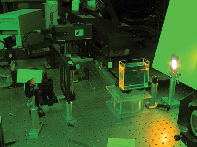
Figure 1. Supercontinuum light generated in liquid from red to white. The experimental setup sends high-intensity laser light (red) through a suitable optical medium (here a container of liquid) that greatly broadens the light’s bandwidth (white light). Courtesy of Robert Alfano.
This article reviews the panoply of applications of the SC.
Nonlinear microscopy to OCT
Recent improvements in SC laser technology have left their mark on biology, studies of condensed matter, chemistry and engineering. There have been notable breakthroughs in high-precision optical frequency and time metrology, ultrahigh capacity wavelength division multiplexing (WDM) communication, and optical coherence tomography (OCT). Because of pulse compression and ultrafast laser pulse generation, the resulting frequency is also possible for combing f − 2f for attosecond pulse generation, extreme accuracy clocks and high-resolution microscopy.
Present-day laser FiOS fiber communication is in the GHz region, though data transmission rates of terabits per second have already been achieved by using a small segment of the SC light spectrum. But many challenges remain, including improving and achieving petabits-per-second operation by using more of the SC bandwidth, reducing the duration of a bit to shorter than a picosecond and increasing the number of coherent wavelengths, which is likely achieved with normal dispersion fiber and series of pulse compressors6.
In the field of nonlinear microscopy, SC lasers are an alternative to Ti:sapphire lasers, adding a new dimension to unravel condensed matter interactions and reveal biological processes, and aid in understanding diseases such as cancer and atherosclerosis, and neurological diseases at the subcellular resolution (Figure 2)4. When used with filters, the light can be selected and generate intrinsic label-free multi-photon fluorescence from tissues’ native biomolecules7.
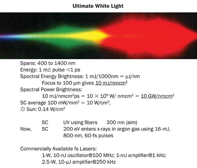
Figure 2. Supercontinuum spectra with various parameters. Courtesy of Robert Alfano.
These light sources have also shown promise for improving OCT8. Early OCT imaging systems had an axial resolution of 10 to 15 µm, relying on a type of diode. The axial resolution depends on the bandwidth of the light source, with a broader bandwidth enabling finer resolution. A short coherence length and a bandwidth broader than any femtosecond laser make SC lasers ideal for achieving high-resolution imaging. The SC light generated in microstructured fibers has been used to produce images of cells with an axial resolution of ~0.5 µm4,8.
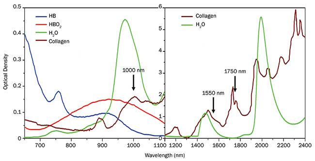
Figure 3. Absorption spectrum for key biological molecules in tissue in the NIR and SWIR. There are four optical windows in this spectrum (600 to 2400 nm) for deeper imaging. NADH: nicotinamide adenine dinucleotide. Courtesy of Robert Alfano.
Broad or selected portions of the SC can be used to selectively excite a particular molecular group or bonds in electronic transitions and vibrational combination and overtones. There are many native molecules in tissues and cells with absorptions from electronic transitions from S0 to S1 and S2. These transitions occur not only in the band of 260 to 600 nm but also in NIR and SWIR regions from 700 to 2400 nm, including the absorptions from water and collagen (Figure 3) and emissions from tryptophan, collagen, flavins and NADH (nicotinamide adenine dinucleotide) (Figure 4), called the building blocks of native biomolecules in tissues. There are four imaging optical windows between 600 to 2400 nm for deeper imaging; the third window, from 1680 to 1800 nm, has the maximum transmittance and minimum absorption, superior to all other windows, and, as such, is called the golden window9,10. Therefore, the application of a SC laser in the SWIR region may optimize the imaging in deeper layers of tissues due to reduced scattering.
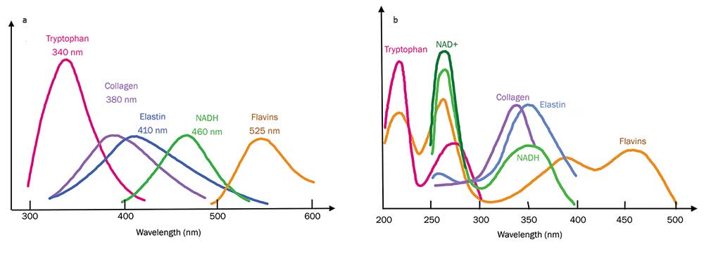
Figure 4. UV to visible fluorescence (a) and absorption spectra (b) of key building blocks of molecules in tissues. See reference 12. Courtesy of Robert Alfano.
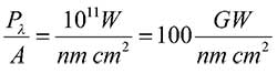
Supercontinuum light can be generated from ~100 femtosecond laser pulses with as low as 10 nJ in energy. Typically, 10 µJ to 10 mJ are used for most applications. With a 1-mJ laser pulse, SC can easily span 1000 nm from visible to NIR region, and the peak spectral energy would be:
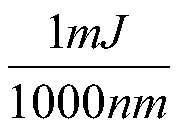
This value will give peak spectral energy of:
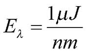
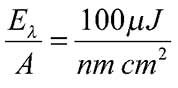
A peak spectral energy flux (irradiation energy per area) of SC light of a beam size of 1 mm is:
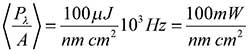
For a 100-fs pulse, a spectral power density is:
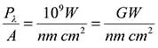
This peak spectral power flux is sufficient to produce a variety of nonlinear optical effects via χ2 and χ3 processes, such as SHG, SRS, 4WM, THG and multiphoton absorption for different spectral filtered bandwidths of 1 to 10 nm at about 1 GW/cm2.
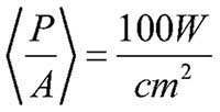
Focusing the SC beam to a 100-µm spot size results in peak spectral power flux at the focal spot of:
for a selected bandwidth of 1 nm at about 100 GW/cm2.
There is enough power to ionize most and create copious nonlinear optical effects with a spectral selected window of a few nanometers. This spectral power of SC light in blue can produce two, three and higher photon absorption transitions in tissue, bacteria and various viruses and can even alter them.
The average spectral brightness of SC for 1-kHz pulse train for 1-mJ pulses is:
The brightness of the SC over entire 1000-nm bandwidth is:
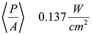
By comparison, the brightness of the sun hitting surface of the earth at 1370 W/m2 is:
Healing light
One can speculate and test whether the SC can be used for therapeutic applications of healing wounds, bruises and burns and welding tissue by exciting electronic and vibrational states of the underlying molecules in tissue and skin. Tissues may be healed and fused with reduced scarring as well11. The underlying mechanism is not clear yet, but it is worth further investigation to test the hypothesis that light may induce angiogenesis and improve healing. Typical irradiance needed for bonding wounds is ~100 mW in the visible to NIR using selected wavelengths.
The upper electronic state excitation by multiphoton absorption (two, three and four photons from the SC) can lead to the rupture of bonds and ionization of polyatomic molecules.
There is the potential for using SC light as an alternative to electrical stimulation for therapeutic treatment of neurological diseases. Typical electrical stimulation of nerves requires metal electrodes inserted into tissue to make contact precisely near the nerves, while optical stimulation can deliver the signal deep into the tissue intact. Supercontinuum NIR laser pulse radiation at 1600 to 2300 nm is potentially possible to stimulate nerves using wavelengths within the optical windows III and IV10.
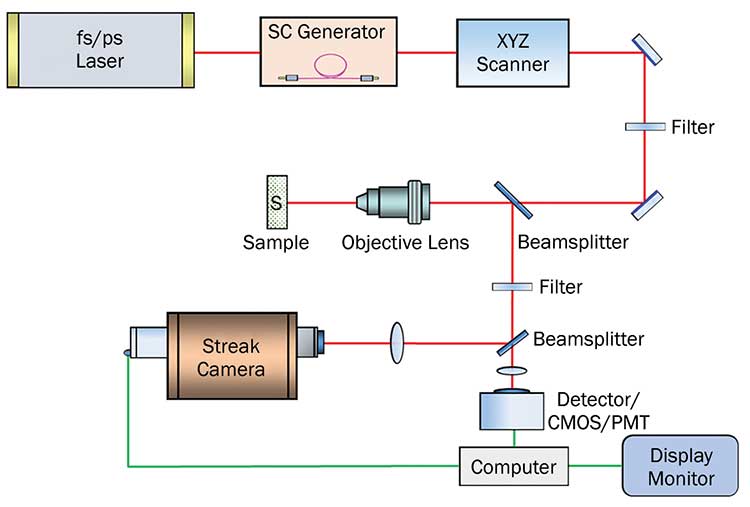
Figure 5. Supercontinuum microscope design. Courtesy of Robert Alfano.
Supercontinuum light sources are now available commercially with average powers up to 50 W and soon will scale even higher to 300 W and above. But there is still a need for smaller, cellphone-sized SC sources for broader applications. The goals are to produce SC optical, NIR and SWIR microscopes for deeper imaging into materials or biological tissues. Images up to 5D tomography may be obtained to form the images of space to get [xyz, t, λ]5. A design of a SC microscope (Figure 5) will combine the spectral and temporal worlds4,7.
Meet the authors
Robert R. Alfano, Ph.D., is a distinguished professor of science and engineering at the City College of New York. He has pioneered many applications of light and photonics technology to study biological, biomedical and condensed matter systems using optical spectroscopy and imaging, including discovering the supercontinuum. Alfano is a fellow of the American Physical Society, the Optical Society and the Institute of Electrical and Electronics Engineers; email: [email protected].
Lingyan Shi, Ph.D., is a research scientist at Columbia University. Her current research focuses on metabolic imaging with stimulated Raman scattering microscopy. She was a research associate studying deep imaging and drug delivery in the brain at the Institute for Ultrafast Spectroscopy and Lasers, the City College of New York, where she received her Ph.D. in biomedical engineering; email: [email protected].
References
1. R.R.Alfano and S.L. Shapiro (1970). Phys Rev Lett, Vol. 24, Issue 12, pp. 584–587 and pp. 592-594, Issue 22, pp. 1219-1222.
2. J.K. Ranka et al. (2000). Visible continuum generation in air–silica microstructure optical fibers with anomalous dispersion at 800 nm. Opt Lett, Vol. 25, Issue 1, pp. 25-27.
3. J.M. Dudley and J.R. Taylor, eds. (2010). Supercontinuum Generation in Optical Fibers. Cambridge, England: Cambridge University Press.
4. R.R. Alfano and L. Shi (2017). US Patent application US 2017/0153435 A1.
5. R.R. Alfano and S.L. Shapiro (1971). Picosecond spectroscopy using the inverse Raman effect. Chem Phys Lett, Vol. 8, Issue 6, pp. 631-633.
6. R.R. Alfano, ed. (2016). The Supercontinuum Laser Source: The Ultimate White Light, 3rd ed. New York: Springer.
7. R.R. Alfano (2016-17). US Patents #9,414,887 B2, Aug. 16 2016 and # 9,561,077 B2, Feb. 7, 2017.
8. N. Nishizawa et al. (2014). Real-time, ultrahigh-resolution, optical coherence tomography with an all-fiber, femtosecond fiber laser continuum at 1.5 µm. Opt Lett, Vol. 29, Issue 24, pp. 2846-2848.
9. L. Shi and R.R. Alfano (2017). Deep imaging in tissue and biomedical materials. Singapore: Pan Stanford Publishing.
10. L. Shi et al. (2016). Transmission in near-infrared optical windows for deep brain imaging. J Biophotonics, Vol. 9, Issue 1-2, pp. 38-43.
11. V. Sriramoju and R.R. Alfano. (2015). In vivo studies of ultrafast near-infrared laser tissue bonding and wound healing. J Biomedical Optics, Vol. 20, Issue 10, pp. 108001.
12. L. Shi et al. (2017). Label-free fluorescence spectroscopy for detecting key biomolecules in brain tissue from a mouse model of Alzheimer’s disease. Sci Rep, 7:2599, doi:10.1038/s41598-017-02673-5.
Light: The Basics
Light may be considered as a sine wave. It is described by key wavelength units measured in nanometer: 440 nm is blue, 530 nm is green and 630 nm is red in the visible light spectrum. The basic equation of light relating the frequency to its wavelength is given by
c=λν,
where c = 3 ×1010 cm/s is the speed of light, λ is the wavelength (nm) and ν is the frequency (Hz).
The relationship between energy and frequency for light from Planck is given by:
E=hν,
where h = 6.626 × 10⁻³⁴ m2kg/s is the Planck constant.
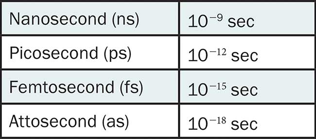
The key unit of light pulses is length measured in:
Self-Phase Modulation and Four-Photon Mixing
Self-phase modulation and four-photon mixing are the key mechanisms behind supercontinuum light generation. Mainly in solids, gases and all normal dispersion fiber, the electronic mechanism of supercontinuum light is related to the distribution and distortion of an electronic cloud of atoms and ionization in intense electric from nonlinear index n2, which is related to the third order susceptibility χ3.
Today, researchers produce supercontinuum light that spans an entire octave or more. An octave of light extends all the way from one frequency to the double of that frequency. The visible spectrum is approximately an octave, and thus the supercontinuum realizes the long-sought goal of white laser light.
The first application of the supercontinuum was studying the dynamics of vibrational excitations in liquids5 and later used by noted chemists, physicists and biologists such as Nobelist Ahmed Zewail. John Hall of the University of Colorado at Boulder and the National Institute of Standards and Technology, along with Theodor Hänsch of the Max Planck Institute for Quantum Optics in Garching, Germany, received their share of the 2005 Nobel Prize in Physics for using such light to achieve extremely accurate time and frequency measurements.
Underlying Physics
When a highly intense light pulse propagates through a medium, the electric field of the pulse can alter the index of refraction, n2, which, in turn, affects the propagation of the pulse. The time-dependent index changes the phase of the wave, which changes the temporal and spectral characteristics as well.
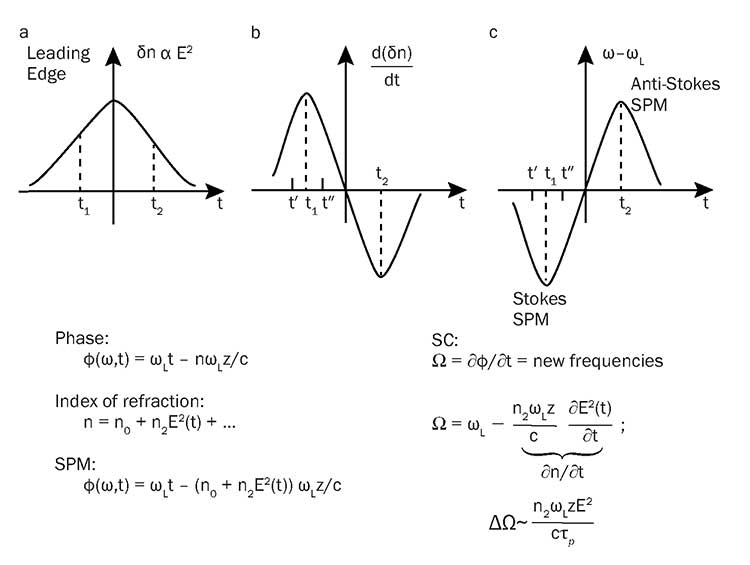
A simple supercontinuum model via self-phase modulation and nonlinear index of refraction. Courtesy of Robert Alfano.
The spectral broadening that occurs is called self-phase modulation or super broadening. There are a number of mechanisms that contribute to the index variation: direct electronic cloud distortion, molecular reorientation, molecular redistribution, vibrational motion of molecules, and multiphoton ionization3,6. Self-phase modulation can be obtained by considering a beam propagating through a medium in which the index of refraction is:
n = n0 + n2E2 + …
where n2 is a nonlinear index of refraction.
Ordinarily, when all but the first term in the expansion is neglected, the beam would be expected to expand with angular divergence. When the nonlinear term is large enough, the critical angle for total reflection at the beam edge will be such that all of the energy is confined to the beam. In other words, spreading of the beam by diffraction is reduced and phase of wave reacts on itself to cause self-effect such as self-phase modulation1. The prior equation can be rewritten as follows in a time-dependent form for a single index affecting mechanism of relaxation time τ:
n(t) = n0 + n2 ∫ exp [−(t-t’)/τ] E2(t’)dt’
The electric field of the incident laser pulse is:
E(t) = E0 [exp (−t2/τP)] cos(ω0t − θ(t) ),
were τp is related to the pulse width at half maximum and the phase changes and induced phase θ(t):
θ(t) = n(t)ωoz/c
The spectral extension in supercontinuum is given by:
Δω ~ n2 E2 ωo Z/τpc
Higher order effects for solitons are neglected here2,3. These do not occur in all normal dispersion fibers6.