When researchers
at Cornell University in Ithaca, N.Y., and the University of Florence in Italy set
out to optimize second-harmonic-generation microscopy, they did so to improve measurements
of neuronal membrane potentials. For this to work, though, they had to overcome
the photodamage that is a byproduct of second-harmonic generation. They succeeded
and uncovered some unexpected effects.
“We were surprised by two aspects
of the photodamage: the significant decrease on slowing the exposure rate and the
cubic intensity dependence,” said Watt W. Webb, research team leader and a
professor of applied physics at Cornell.
Unlike electrode-based methods, second-harmonic-generation
microscopy can measure membrane potential from many neurons at the same time, and
it does so with micron and millisecond resolution. What’s more, the signal
originates only from dye molecules that are properly ordered in the plasma membrane,
and the technique allows deep-tissue, high-resolution imaging.
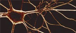
By tweaking their setup, researchers imaged neurons of the sea slug
Aplysia with second-harmonic generation but without photodamage. They used a membrane
potential sensitive second-harmonic-generating dye at the right concentration and
illuminated it with the proper laser power. The electrical activity of the neurons
changed the intensity of the emitted second-harmonic signal and allowed them to
optically record neural activity in real time. This image measures about 150 x 450
μm. Courtesy of L. Sacconi and D.A. Dombeck.
Because the second-harmonic-generation
signals that track voltage responses are small, achieving an acceptable signal-to-noise
ratio typically requires intense illumination and/or high dye concentrations.
One solution is to average multiple line scans, but that doesn’t work in some
situations.
According to the researchers, photodamage
probably results from multiphoton excitation that accompanies second-harmonic generation.
The researchers set out to find a recipe for conditions that maximize second-harmonic generation while minimizing photodamage. They did this by comparing the resting potential of Aplysia neurons as measured
by an electrode before, during and after illumination under various conditions.
They measured photodamage by its effect
on resting membrane potential during line- and image-scanning modes, varying laser
illumination from 0 to 100 mW and dye dose concentration from 0 to 25 μM. They
also used OxyFluor, an oxygen scavenging enzyme, from Oxyrase Inc. of Mansfield,
Ohio, to examine the dependence of photodamage on oxygen. Webb noted that varying
all these parameters involved quite a bit of work.
For staining, the scientists used FM4-64
dye. The imaging system consisted of a fiber laser from Fianium Inc. of Eugene,
Ore., coupled to a Bio-Rad scan box and an Olympus microscope, with Hamamatsu gallium-arsenide-phosphide
photomultipliers sitting behind an optical filter for a detector.
These measurements showed that the
photodamage increased linearly with the dye concentration but grew with the cube
of laser intensity. The latter might be due to a third-order photochemical process,
such as three-photon dye excitation, but this has yet to be pinned down. Adding
the appropriate antioxidants before the laser scan lessened photodamage.
Applying these results, the researchers
used second-harmonic-generation microscopy to record membrane action potentials
in a single trial, a technique that could prove useful for real-time investigations.
They also recorded action potentials along neuron neurites, although they had to
cut laser power approximately in half to do this.
These findings help point the way to
the researchers’ ultimate goal of optimizing optical imaging of fast changes
in membrane potential anywhere in a neural system. Webb said that achieving this
might become easier with advances in technology. “There is always the hope
of developing better dyes,” he said.
PNAS, Feb. 28, 2006, pp. 3124-3129.