Dr. Stuart K. Bisland, Princess Margaret Hospital/University Health Network
Canada has demonstrated its commitment to building a sustainable
future for biophotonics through education, research and industrial development.
Numerous initiatives exist, at federal and provincial levels, whose mandate is to
establish a firm foothold for biophotonics within the global arena. For example,
the Advanced Biophotonics Consortium (ABC), established in April 2003, comprises
life sciences and photonics organizations and universities and colleges. Its goal
is to create a globally competitive entity with a focus on biophotonics.
ABC, in partnership with Carleton University in
Ottawa and the National Research Council of Canada, also established the Canadian
Photonics Fabrication Centre in Ottawa that aims to develop innovative photonic
devices specific to, among other things, biomedicine. The research council also
partnered with Carleton, along with the University of Ottawa, in 1998 to establish
a training initiative called Vitesse Reskilling, whose mandate includes maximizing
the transfer of skills derived from photonics into other areas, predominantly biophotonics.
Since 1993, Vitesse Reskilling has run a series of biophotonics symposia in alliance
with the NATO Advanced Study Institute, which is aimed at consolidating the current
and future directives for biophotonics in Canada and the rest of the world.
An important cornerstone of the Canadian
commitment to biophotonics now is education, which had been less of a priority,
especially for undergraduate and even younger students. Biophotonics has not been
featured greatly in Canadian schools, colleges or universities but, rather, confined
within research facilities or teaching hospitals.
No undergraduate biophotonics course
is offered in any Toronto academic institute. Attitudes are changing, however.
Within the past couple of years, Niagara College in Welland, Ontario, and Algonquin
College in Ottawa began to offer a bachelor’s degree in photonics, including
a biophotonics component. Comparable courses are now available at a number of universities
in and around Canada, including Simon Fraser University in Burnaby, British Columbia,
and McMaster University in Hamilton, Ontario.
I founded a new initiative called SABRE
(Strategies for Advancing Biophotonics Research and Education) to improve the
teaching of biophotonics in schools as well as the general awareness of the subject
in the public sector.
Exposing schoolchildren and undergraduates
to the exciting science of biophotonics and career opportunities that exist therein
is fundamental to the continued accrual of students at the college and university
levels and, ultimately, to the continued expansion and success of biophotonics.
PDT research
Research in Canada includes fluorescence lifetime
imaging, fluorescence and diffuse reflectance spectroscopic techniques, interferometry
and optical coherence tomography, Raman spectroscopy and optoacoustics. One particularly
strong area of research, resulting in commercialization, in some cases, is photodynamic
therapy (PDT).
Quadra Logic Technologies Inc. of Vancouver,
British Columbia, has commercialized the Photofrin photosensitizer and Visudyne
therapy, which is used to treat classic subfoveal wet-form age-related macular degeneration.
PDT research continues throughout the
country. At the BC Cancer Research Centre in Vancouver, researchers Mladen Korbelik,
Stephen Lam and Calum MacAulay are studying the role of tumor immunology in PDT,
fluorescence imaging and the treatment of cancers.
A large part of our research at the
Princess Margaret Hospital/University Health Network centers on PDT. Dr. John Trachtenberg,
Brian C. Wilson, Dr. Paul J. Muller and Fred W. Hetzel are leading two ongoing
clinical trials for PDT treatment of recurrent prostate cancer and glioma-derived
brain tumors. In the laboratory, we recently demonstrated PDT to treat primary and
secondary bone cancers, and preparation is under way for Phase I clinical trials
involving the treatment of late-stage bony metastases. This work has led to applications
unrelated to cancer, including treatment of bacterial infections within bone and
studies to examine the underlying mechanisms of PDT effects on bone development.1
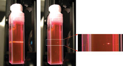
Figure 1. A dye in a cuvette is excited using one-photon (left) and
two-photon (right) fluorescence excitation. Two-photon excitation provides a three-dimensionally
localized (at the focal point) effect for PDT as compared with one-photon excitation,
which excites throughout the laser beam path.
Another type of PDT treatment is being
researched by David T. Cramb’s group in the department of chemistry at the
University of Calgary in Alberta, by our group in Toronto and by Melanie C.W. Campbell’s
group in the department of physics at the University of Waterloo in Ontario. These
teams are collaborating on two-photon strategies for PDT treatment inside the eye.
Aliaksandr Karotki, a postdoctoral fellow with Wilson in Toronto, is leading the
study and has engineered a femtosecond laser scanning confocal ophthalmoscope that
allows two-photon PDT at the back of the eye. It is believed that two-photon PDT
will be able to treat very precise, clearly delineated volumes within deep tissue
and will abrogate the collateral damage associated with single-photon PDT
(Figures 1 and 2).
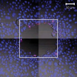
Figure 2. An area of monolayer endothelial cells is targeted for two-photon PDT. The irradiated
area is inside the white square. Staining with Hoechst 33258 (blue) and Sytox (red)
stains only cells with compromised membrane.
Cramb also is doing research that uses
two-photon excitation fluorescence cross-correlation spectroscopy to decipher receptor-ligand
interactions and signal transduction pathways. He recently used quantum dots to
reduce spectral overlap when multiplexing two or more signals.2
Imaging techniques
In much of our preclinical in vivo work, we use
bioluminescence to follow the response and progression of disease in animals before
and after treatment. Alex Vitkin, a senior scientist at Ontario Cancer Institute
& University Health Network at Princess Margaret Hospital and his postdoctoral
fellow Victor Yang have developed an imaging method that employs an endoscopic probe
that allows Doppler measurements of microvascular blood flow using optical coherence
tomography (OCT). This has led to a clinical trial using OCT to examine altered
microvascular blood flow in patients who have Barrett’s esophagus through
a collaboration between our biophotonics group at University Health Network and
the gastrointestinal endoscopy team at St. Michael’s Hospital in Toronto (Figure
3).3 The evolution of OCT from being primarily a structural morphological imaging
modality to one that provides functional information holds great promise and is
an intense area of research.
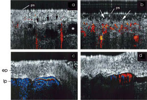
Figure 3. A clinical trial using color
Doppler OCT reveals altered microcirculation patterns in patients who have Barrett’s
esophagus (a and b) and enlarged esophageal veins (c and d). In Figure a mucosal
glands (asterisk) are clearly delineated within the epithelial (ep) Barrett’s
esophagus, which interfaces with the underlying lamina propria (lp, arrows). Superficial
glandular structures are seen in Figure b (arrows) with microvasculature close
to the surface.?Ps indicates the external surface of the OCT imaging tip.
Light-induced fluorescence endoscopy
is another imaging technique that has been commercialized. BC Cancer Research Centre’s
Lam, with Bruno Jaggi, worked with technologies for imaging autofluorescence associated
with precancerous and cancerous lesions in the lung and gastrointestinal tract.
They formed Xillix Technologies Corp. in 1989, and in June 2005, the Xillix Life
was approved by the FDA as an early detection/diagnostic imaging tool for lung cancer.
Assessing burn recovery
Biomedical Photometrics Inc. of Waterloo, Ontario,
founded in 1994 by A.E. Dixon, has developed laser scanning instrumentation that
combines patented wide-field fluorescence confocal technology with automated functionality.
One fast-growing area within biophotonics
is that involving macro/microscopic and spectroscopic imaging techniques and the
development of related biomarkers and biosensors. Lorenzo Leonardi’s team
at the National Research Council’s Institute for Biodiagnostics in Winnipeg,
Manitoba, is developing a near-infrared spectroscopic platform to assess oxygenation
and blood flow within burn-injured tissues. Clinical trials are being conducted
at Sunnybrook Hospital Burn Centre in Toronto.
Predicting and monitoring the recovery
of burn tissue is fundamental to planning treatment strategies and producing the
best outcome for patients. The noninvasive, comparatively inexpensive cost of near-IR
spectroscopy should encourage its use in hospitals.
Recent work by Ulrich Krull and Paul
Piunno at the University of Toronto involves fiber optic-based nucleic acid biosensors
aimed at screening for genetic markers of disease. The biosensors consist of oligonucleotide
probes covalently bound to silica fibers onto which fluorescently labeled target
nucleic acid sequences can hybridize. Fluorescence detection is conducted via the
same fiber as internal reflectance. The researchers have demonstrated clinical utility
by targeting the mutated survival of motor neuron gene implicated in spinal muscular
atrophy.4
Similar optically based sensors for
recognizing single nucleotide polymorphisms in DNA are being developed by Denis
Boudreau’s group at Université Laval in Quebec. The group continues to
refine the development of optical biosensors for rapid and quantitative genetic
testing that will, no doubt, have a significant impact in how biodiagnostics is
conducted in the future.
Nanobiophotonics
Improved microscope technology has allowed light
to target nanoscale objects. Nanoparticles can be used to interrogate DNA/protein
interactions, intracellular transport and transduction signaling. Although in its
infancy, recent work by Warren Chan at the Institute of Biomaterials and Biomedical
Engineering in Toronto exploits the unique tunable properties and stability of
quantum dots for biomolecular fingerprinting of DNA in disease employing Förster
resonance energy transfer (FRET), whole-body imaging of diseased sites and drug
delivery.
Chan and others are working to provide
biocompatible, nontoxic quantum dots emitting in the near-IR spectral region for
intravascular and/or localized deep-tissue imaging in vivo. Studies by Steven Dunn
of the department of biochemistry, University of Western Ontario in London, have
employed a unique FRET paradigm known as time-resolved single-molecule FRET. Based
on the information that FRET provides on intramolecular distances, Dunn has plotted
these distances over time to chart the relative structural rotations of subunits
of F0F1-ATP synthase complex during ATP formation.
This study clearly demonstrates the
application of optical approaches to noninvasively measure conformational dynamics
of important biomolecules, a technology that will undoubtedly prove invaluable
for understanding changes in biomolecular conformation in relation to specific
intracellular transduction pathways.
Fluorescence microscopy
Fluorescence microscopy also can be used to measure
the rate of transport or migration of target proteins within cells using a technique
called fluorescence recovery after photobleaching (FRAP). Areas within a cell containing
a fluorescently tagged protein can be photobleached using two-photon microscopy,
and the time to recover the fluorescence is measured. Information regarding the
role of target proteins within cells can be gleaned for following different treatments
and for differing cell types whether cancerous or normal. Work by Kevin Hadley,
a doctoral student at the University of Toronto, demonstrates the use of FRAP with
HeLa cells that constitutively express heat shock protein (HSP)-70 labeled with
GFP (Figure 4).
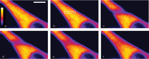
Figure 4. When researchers performed FRAP on a cell with GFP-labeled heat shock protein,
the nucleus (lower right) showed no fluorescence. The images show the cell before
FRAP (a), with a box designating the target area for FRAP (b), immediately after
photobleaching (c), after 75 ms of fluorescence recovery (d), after 300 ms (e) and
after 1000 ms (f). The scale bar is 10 μm.
Plans are under way to consolidate
expertise in biophotonics worldwide under a single Web site called biophotonics world.org.
A new generation of investigators are eager to cross the lines drawn between differing
scientific disciplines, bringing engineers together with biologists, physicists
and clinicians to strengthen the impact of biophotonics in many sectors.
New multimodality approaches in medicine
are improving detection and therapy options for patients. Also, strategies are being
investigated for integrating optical technologies with imaging modalities in clinical
use, such as combining fluorescence imaging with MRI and CT. I think one important
technological development in this regard will be clinical integration of cameras
that allow easy imaging and quantification of hyperspectral fluorescence signals
from biological tissues in situ and in vivo.
Meet the author
Stuart K. Bisland is a scientist at Princess Margaret
Hospital/University Health Network, Toronto; e-mail: [email protected].
References
1. S.K. Bisland et al (January 2006). Pre-clinical
in vitro and in vivo studies to examine the potential use of photodynamic therapy
in the treatment of osteomyelitis. Photochem Photobiol SCI, pp. 31-38.
2. J.L. Swift et al (Feb. 15, 2006).
A two-photon excitation fluorescence cross-correlation assay for a model ligand-receptor
binding system using quantum dots. Biophys J, pp. 1396-1410.
3. V.X. Yang et al (June 2005). Endoscopic
Doppler optical coherence tomography in the human GI tract: initial experience.
Gastrointest Endosc, pp. 879-890.
4. J.H. Watterson et al (Jan. 23, 2004).
Rapid detection of single nucleotide polymorphisms associated with spinal muscular
atrophy by use of a reusable fibre-optic biosensor. NUCLEIC ACIDS RES, pp.
e18.