
Lens-Free Microscopes Offer Real-Time Bio Imaging
Jan Provoost, IMEC
A totally different type of microscope will be much smaller and less complex than traditional microscopes, but more powerful.
Two years ago, a customer handed Richard Stahl and the imec team a challenge: one year to develop a microscope that could help monitor the growth of stem-cell cultures. The customer was already building a prototype using
a phase-contrast microscope, but Stahl decided to try lens-free microscopy instead.
“A lens-free replacement would lower the cost and complexity of their application,” he said. “And it would also allow them to capture a much larger field of view, the surface that you see with the microscope. And this in one shot, without mechanical scanning and image stitching. But of course – and this was our challenge – its resolution would have to be as good as that of the phase-contrast microscope.
“In retrospect, I don’t think they believed we could do it.”
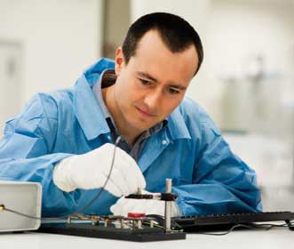
A team of researchers at imec has developed a lens-free
microscope that can compete with phase-contrast microscopy. Photo
courtesy of imec.
The principle of lens-free imaging was first formulated by the Hungarian-born engineer Dennis Gabor in the late 1940s. For perfect imaging, he reasoned, you need not only the amplitude of the diffracted light (which gives you gray-scale 2-D pictures), but also phase information (which gives you thickness and depth information). He proposed to capture – on a photographic plate – the interference pattern between the light that illuminates an object and the light diffracted by the object. In this pattern, all information is available to reconstruct an image of the object. This achievement, along with his other work, earned Gabor the 1971 Nobel Prize for physics.
A decade ago, with the advent of powerful CMOS imagers, the photographic plate could be replaced with a fast, high-resolution digital image sensor. And as computers became powerful enough to do the required calculations in a reasonable time, it became possible to convert the captured holograms into useful images.
Developed at several labs around the world, lens-free holographic microscopy can now yield images with a resolution well below 2 μm over a large field of view (e.g., 5 × 5 mm). Moreover, the image reconstruction and interpretation are completely digital, using powerful yet affordable computers.
Geert Vanmeerbeeck shares an office with Stahl. Together, they’ve been working on lens-free microscopy for almost two years.
“Given our track record with imaging and its integration,” Vanmeerbeeck said, “it was only natural for us to start working on lens-free microscopes.
“But we didn’t set out with the intention of adding yet another flavor or publishing yet another flight of papers, though. We want to integrate and miniaturize the technology to make it applicable to many more applications, some of which will be real breakthroughs.”
Looking for the ghost buster
“One year into the stem-cell project,” Stahl said, “we had reached a resolution of approximately 2 µm, but still with a lot of noise. Just good enough to continue the project, but we would have to do better.”
The competition – the phase-contrast microscope – had been developed specifically to look at living biological material, which is mostly transparent. With traditional microscopes, laboratory technicians first have to stain the samples to reveal structures. But such staining changes and sometimes even kills the samples, making the method unusable for many applications. In a phase-contrast microscope, light travels through a transparent, living sample. The image is made from the phase changes that the light undergoes in the sample.
Lens-free holography also images transparent material. But the recording suffers from an inherent side effect called twin image distortion. Because an image sensor essentially captures only the intensity of light, all phase information gets lost, so that the computed reconstructed image shows the object in focus, along with a fainter ghost of the object at twice the focal distance. The resulting picture is … blurry.
“To eliminate the ghost object in the computation, you need more information about the object under the microscope,” Vanmeerbeeck said. “The best way to do that is to take several slightly different pictures and combine these to retrieve the phase information. This is called ‘iterative phase retrieval.’ ”
Taking these different pictures could be done by changing the distance between the sample and the image sensor, for example. “But to do that,” Vanmeerbeeck said, “we would need mechanical movements, bringing back in all sorts of hardware alignment issues that we had set out to eliminate with our lens-free setup.
“What we do instead is take the same picture several times, but each time using a coherent light source with a slightly different wavelength. This way, we have different pictures of the same scene, but without movement. Using a combination of these images, we can eliminate the blurry ghost effect and get sharp microscope images.”
“So last May,” Stahl added, “we were able to show a setup for stem-cell monitoring with a 1.3-µm resolution, similar to the resolution of a phase-contrast microscope with 10× objective. Our system, however, has a field of view that is 14 times larger.”
To catch a heart cell contracting
With a working lens-free solution for one application, the “lens-free team” set out to diversify and see how it could adapt its first experiences to other applications.
“A fascinating example is imaging the contractions of heart cells,” Stahl said. “It’s currently a hot topic in drug discovery to try and test potential new drugs on cells and tissues grown in the lab, instead of using live animals. And you certainly want to test [whether] a new drug [interferes] with the contraction of the heart, or even blocks it altogether.”
To monitor contracting heart cells, you look at the changing shape of a relevant number of cells. You need a large field of view, but you don’t necessarily need to see all the details of the cells. And to see the changes in contraction speed, which may be subtle, you need a high imaging speed, higher than other microscopes provide. Stahl and Vanmeerbeeck have already tested imaging at a few hundred frames per second, and they think this application may require even higher speeds.
“Heart cells normally contract when they receive a high-enough electrical stimulus,” Stahl said. “But drugs can interfere with that reaction. So next to imaging the contraction, we also want to measure the relation with the electrical activity in the cells.”
To do that, the researchers grow the cells on a chip with an array of microelectrodes. Imec has pioneered such chips with a biocompatible surface with hundreds of ultrasmall nails – microelectrodes that can monitor and stimulate individual cells that grow on top.
“These surfaces are not transparent, so you can’t shine light through a sample and capture the result at the other side,” Stahl said. “Instead, we use the light that reflects from the cells and the surface below the cells, a technique that is called reflective holography. The way we do that is really innovative.”
Seeing inside the chip
To study cell cultures growing in a petri dish, you can use a relatively small microscope, but there is no need to make the microscope … well, microscopic.
“Our colleagues in the life science department are looking to develop chips that analyze biological material; for example, looking for specific cells, bacteria or DNA sequences,” Vanmeerbeeck said. “On those chips, you need microfluidic channels, and chambers for chemical reactions. But you also need a microscope. And you need it small, cheap and without mechanically moving parts. So that would make our lens-free microscope the ideal candidate. That is, if you are able to microsize all the components.”
That is exactly what Dries Vercruysse is looking into. Vercruysse is a researcher in imec’s biophotonics group, looking to adapt the lens-free microscope for use inside chips.
“In these applications,” he said, “we are not so much concerned with a large field of view. We want to look at one cell or bacterium at a time and determine its type and characteristics. So we need to see it as sharp as possible. The industry fabricates ever-better imagers with smaller pixels, but today we’re still talking about pixel sizes of 1.5 to 2 µm. And that pixel size essentially limits the resolution of our lens-free microscopes, unless we pull a trick.”
Vercruysse’s solution is to magnify the beam of light before it falls on the imager, again without using lenses. He illuminates the sample through a microscopic pinhole. At the back of the hole, the light beam forms a widening cone, shining first through the sample and eventually falling on the image sensor. “By varying the distance between the pinhole and the sample, we can vary the magnification without using lenses,” he said.
One driver of imec’s bioelectronics R&D is the iLab, a laboratory on a chip that the research center is developing together with Johns Hopkins University. The iLab is conceived as a breakthrough diagnostic tool that will incorporate a host of nontrivial diagnostic tests. It will be able to do a viral-load count in a drop of blood, for example. And the lens-free microscope will be one of the enabling tools inside the iLab.
Let a thousand microscopes bloom
The researchers currently are designing a lens-free microscope that fits on a chip; they also are already thinking about a chip that would hold one thousand such microscopes. At the start of 2014, imec’s Liesbet Lagae was awarded a prestigious grant from the European Research Council to further develop a cell-sorting chip. Lagae is imec’s R&D manager for life science technologies and a professor of nanobiophysics at the University of Leuven.
Her cell sorter is a platform for ultrafast inspection and sorting of cells. It can be used, for example, to distinguish between cancer cells and normal cells in tissue studies or biopsies, or even directly in a blood sample. This way, cancer metastasis can be detected early on, improving the patient’s outlook considerably.
“In its most powerful specifications, the cell sorter will have a thousand channels, each inspecting a thousand cells per second, determining if they are malignant and mechanically sorting them into separate chambers,” Vercruysse said. “Each channel will have a lens-free microscope that will record a hologram of a cell, reconstruct and analyze the image, and make a decision in one millisecond.”
“The cell sorter sets us a number of challenges that go beyond what anybody has done today with lens-free microscopy,” Vanmeerbeeck said. “Take the speed of imaging, which is an order of magnitude above what we do now. Then we have to distribute the light source over a thousand microscopes. And will we use a thousand small imagers, or one big shared imager? At this moment, we’re modeling the various options to find the best way to build our microscopes into the cell sorter.”
For Stahl, designing these lens-free microscopes is fascinating work: “As we find ways to make them smaller, or improve the resolution, more people get interested, and more applications become possible,” he said. “But it also works the other way around. As partners challenge us to try and match our microscopes into their applications, we work on new approaches. But what is really unique here, with the expertise and tools we have at imec, is that we are also able to integrate and implement our ideas into working prototypes.”
Meet the author
Jan Provoost is science editor at imec in Leuven, Belgium; email: [email protected].
Published: September 2014