Daniel C. McCarthy, Senior Editor
Scientific lasers are in the foreground as an applications market
but, in the lab, they must deliver the functionality, reliability and ease of use
to remain in the background as tools.
Laser manufacturers take note: A nonscientific survey indicates that scientists want a highly tunable
turnkey laser with configurable pulse widths and repetition rates. It should be
easily mode-locked, have good shot-to-shot and long-term stability, be ready to
use with the flip of a switch and cost little more than an optical microscope.
“Yes, I want a laser that puts
out all wavelengths and that operates from continuous-wave to attosecond pulses
and that costs next to nothing,” said Richard Zare, director and eponym of
the Zarelab at Stanford University in Stanford, Calif. And yes, he was speaking
tongue-in-cheek, but he was much more sincere when he said, “Without lasers,
most of my research wouldn’t happen.”
That applies to research beyond Stanford,
too, limited though lasers are when compared with Zare’s ideal. In fact, scientific
laser markets fared well while other revenue streams were drying up for technology
vendors. Several suppliers reported that sales of these devices had remained strong
while other laser products had declined. Coherent Inc. in Santa Clara, Calif., reported
that its scientific laser sales actually increased 20 percent amid an otherwise
sluggish laser market.
Driving those sales were academic researchers
as well as analytical instrument manufacturers and integrators, such as Becton Dickinson
and Jobin Yvon Horiba. Spectroscopy — including pump probe, fluorescence,
Raman and anti-Stokes Raman techniques — remains the most prevalent application,
but the hottest growth has been in multiphoton excitation, a field that one laser
supplier described as halfway between a commercial and a scientific market.
“The more exposure end users
get to multiphoton techniques, the bigger the market will get,” said Paul
Ginouves, business development manager for Coherent. “Multiphoton excitation
is in the same kind of place that flow cytometry was 20 years ago.”
Although still in an embryonic stage,
multiphoton research is evolving into a hybrid field that encompasses physics, chemistry
and biology. “So researchers, although they’re not laser jockeys, want
to get the maximum functionality when they plunk down $250,000,” Ginouves
said. “That drives them toward ultrafast technology because these lasers have
the broadest tunability and functionality.”
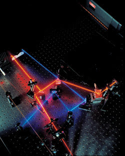
Typically, applications drive the research. Ultrafast Ti:sapphire lasers, however, continue to enable new
and unanticipated experiments. Courtesy of Coherent Inc.
But functionality does not equal complexity.
Most multiphoton users are biologists and chemists who prefer to focus on the experiment
rather than on its tools. “Physicists don’t mind as much because they
have fun with lasers and they don’t mind tweaking them once and a while, but
chemists and biologists expect more ease of use,” said Arndt Kruger, director
of marketing for the industrial and scientific business unit of Spectra-Physics
in Mountain View, Calif.
Lasers on the brain
When Marvin Minsky developed the first confocal
microscope in the 1950s, he speculated on its potential for mapping interconnections
of live brain cells. The application never coalesced, in part because the light
sources available at the time were noncoherent and comparatively dim, which prolonged
scanning times. Instead, confocal microscopy waited nearly 40 years for lasers that
were compact and powerful enough to make the technique practical. In the relatively
short time since then, multiphoton microscopy — evolved from confocal techniques
— has realized Minsky’s dream.
Both multiphoton and confocal microscopes
rely on laser light to excite fluorescence in a sample, but multiphoton technology
uses two or more infrared photons instead of confocal’s single visible or
ultraviolet photon. Also, infrared radiation not only penetrates deeper into tissue,
but also incurs less damage to cells and tissue outside of the focal plane, which
enables extended three-dimensional imaging of living tissue.
Multiphoton — specifically two-photon
— techniques underlie neurobiologist Steve Potter’s attempts over the
last three years to teach rat neurons a lesson. Potter affixes neurons taken from
embryos into electrode-embedded petri dishes that maintain two-way communication
between the cells and a computer. He hopes that if he can coax his simulated animals
— called “animats” — to learn, he can make movies demonstrating
how brain cells make connections that enable learning. The plot of his movies will
unfold under a multiphoton microscope driven by a Ti:sapphire laser.
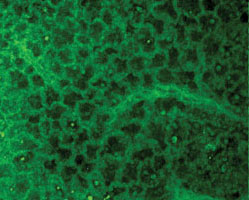
A Ti:sapphire emitting 150-fs pulses at 850
nm enabled a multiphoton microscope to capture this image 100 to 150 μm inside
a mouse’s lung and allowed biologist A.M. Gurney to examine samples. Confocal
scanning microscopy, however, can reach depths of only about 40 μm. Courtesy
of Strathclyde University.
Within the context of his work, Potter
regards Ti:sapphire lasers from Coherent and Spectra-Physics to be interchangeable,
but his primary criterion for selecting the laser is how many milliwatts of power
it can provide at various wavelengths. Hence, when he recently moved his research
from the California Institute of Technology in Pasadena to the Georgia Institute
of Technology in Atlanta, he obtained a 10-W Nd:YAG laser to pump his Ti:sapphire.
The additional power will help the ultrafast laser tune from 700 to 1000 nm. The
more wavelengths he can elicit from his system, the more fluorophores he can apply.
In fact, laser selection for most fluorescence-based
research stems from the fluorophores needed to “see the biology,” as
one researcher put it. The fluorophore determines the wavelength of the excitation
source and, to some extent, the power required.
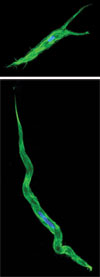
The biology determines
the fluorophore, and the fluorophore determines the laser wavelength. Tunability,
therefore, is crucial, particularly when imaging with two or more fluorophores,
such as when Karen McCloskey captured this multiphoton image of an interstitial
cell from a guinea-pig bladder labeled with actin (green) and DAPI (blue). Multiphoton
excitation can also enable biologists to obtain two or more fluorescent signals
with one measurementbecause it doesn’t bleach out a sample’s fluorophores
with one shot. Courtesy of Strathclyde University.
For instance, to image calcium within
sample cells, you would probably look for a source emitting 800 to 820 nm in 100-
to 200-fs pulses, said John Girkin, associate director of the Institute of Photonics
at Strathclyde University in Glasgow, UK. However, he added, another researcher
in the same lab might be capturing images with green fluorescent proteins, which
would require the source to operate at around 920 nm as well.
Their tunability and pulse widths,
therefore, make Ti:sapphire lasers suitable for a broader range of applications.
It also makes them more expensive. Girkin purchased four multiphoton microscopes
from Bio-Rad Microscience in Hemel Hempstead, UK, in the last 18 months. The systems
are based on Ti:sapphires that, by themselves, cost in the neighborhood of $100,000.
Consequently, Strathclyde’s institute
is working on a new generation of sources that are not tunable but that operate
at one wavelength with a low component cost — about $5000.
“This means that a personal multiphoton
imaging system is possible,” Girkin said. “If one then wished to look
at other samples later, one would just purchase a second laser source at 920 nm.”
Such sources provide a tuning range
of only about 30 nm. But by gradually adding systems, full spectral coverage would
become available and cost-effective in comparison with a single Ti:sapphire.
At the University of Virginia’s
Keck Center for Cellular Imaging in Charlottesville, Ammasi Periasamy is writing
grant proposals to obtain more multiphoton systems. In the last four months, he
has added a Ti:sapphire laser to his lab’s arsenal of two. Integrated into
multiphoton microscopes, the lasers enable his group to track the movement of cells
in frog embryos. The researchers are also studying calcium emission in neurons,
which could help them get a better handle on Alzheimer’s disease.
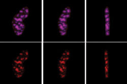
Higher pump powers increase the tuning range
of Ti:sapphires and the options for available fluorophores. In the case of this
fluorescent resonance energy transfer image, a 10-W pump enabled a Ti:sapphire
laser tuned to 820 nm to excite the donor molecules and, tuned to 950 nm, to excite
the acceptor molecules. The images show a three-dimensional reconstruction of C/EBPα
proteins expressed with CFP-dsRED in GHFT1-5 living cell nucleus. Courtesy of the
University of Virginia’s Keck Center for Cellular Imaging.
Parallel to this research, the group
is monitoring several analytes together to compare their functional activity. Labeling
and imaging three probes simultaneously is possible with confocal scanning microscopy,
but it would require three measurements.
Multiphoton excitation enables Periasamy
to obtain all three signals in one measurement because it doesn’t bleach out
a sample’s fluorophores with one shot.
Unfortunately, there is no solution
as simple as this for reducing the demand for more multiphoton instrumentation.
“Once you set up a system for
a particular experiment, you can’t use it for something else,” Periasamy
explained.
Ultrafast CARS
Where multiphoton fluorescence studies tie up
one laser, coherent anti-Stokes Raman scattering — better known as CARS —
ties up two or more. And that’s just the Ti:sapphire instruments. The technique
tunes the frequency difference of two or more laser beams to match a molecular resonance
within the sample. When excited by laser pulses, targeted molecules emit intense
Raman scattering toward the blue range of the spectrum (i.e., clear from the excitation
signal).
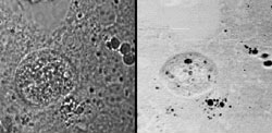
Coherent anti-Stokes Raman spectroscopy helped determine whether these spherical vesicles
in mice hepatocytes were composed of water or lipids. The technique tunes two ultrafast
lasers to correspond with a target molecule’s vibrational frequency. This
eliminates the need for fluorophores but requires pulse widths on the order of 1
or 2 ps. Courtesy of Pacific Northwest National Laboratory.
The technique provides an alternative
for live-cell imaging without the use of fluorescent labels, but it exchanges the
single laser and complex detection system of Raman and infrared-based microscopes
for a complex laser system and a simple single-channel detector.
“As you increase the number of
lasers, the probability of getting everything working together at the same time
becomes more and more remote,” said Gary Holtom, a chief scientist at Pacific
Northwest National Laboratory in Richland, Wash.
Holtom’s coherent Raman setup
includes two Ti:sapphire lasers — an oscillator and an amplifier — an
argon-ion laser pump and an optical parametric amplifier (OPA). Add an array of
beamsplitters, neutral density wheels, bandpass filters and other components, and
you begin to understand Holtom’s observation: “You have to have a commitment
to these lasers to own one. Our total commercial investment is about $400,000. This
is not an experiment you undertake lightly. To make a long story short, there’s
way too much stuff on the table.”
Argon-ion laser technology has continued to outlive its obituaries: Many scientists are loath
to put their old workhorses to pasture. But 20-mW solid-state lasers emitting at
488 nm (shown) may be followed soon by 200-mW versions, which would deliver outputs
comparable with water-cooled argon-ions while diminishing the draw on power consumption
and bench space. Courtesy of Coherent Inc.
Like fluorophore-based applications,
the tunability of Ti:sapphire lasers is an advantage in anti-Stokes Raman techniques.
But unlike straightforward fluorescence microscopy, in which the fluorophore determines
laser wavelength, coherent anti-Stokes Raman scattering techniques tune the laser
according to the target molecule’s vibrational frequency. Also, a laser’s
peak power and spectral bandwidth play a critical role.
Holtom aims for the highest peak power
that he can get to elicit more intense signals. But that requires condensing the
pulse widths to increase their power. The problem is that, when you shorten a pulse,
you expand its bandwidth, which excites signals from additional molecules in the
sample.
As a rule of thumb, transfer-limited
pulses from a Ti:sapphire are 10 to 100 fs in width. That translates into signals
with 100 to more than 1000 wave numbers, which washes out all the useful molecular
information that Holtom is after. Gires-Tournois interferometers integrated in the
laser cavity enable pulses anywhere from 3 to 100 ps.
“So I’m in kind of a no-man’s
land,” Holtom said. “One picosecond just happens to correspond to the
spectral width I’d like to get to. That’s where most molecules happen
to have spectral widths of 10 to 30 wave numbers.”
His former colleague Sunney Xie may
have found a solution to this dilemma. Now at Harvard University in Cambridge, Mass.,
Xie has used two synchronized 5-ps pulse trains to yield better images.
The difficulty is controlling jitter.
The 5-ps pulse trains tolerate no more than 0.5 ps of jitter. A good compromise,
as both Holtom and Xie said, is 2-ps pulse widths, which tolerate less than 200
fs of jitter.
Recently, Xie’s group collaborated
with Jun Ye’s group at JILA in Boulder, Colo., on an optimal laser source
for coherent anti-Stokes Raman scattering microscopy that reduces jitter between
two Ti:sapphire lasers to 20 fs using specialized feedback electronics.
If commercialized, it would provide
the higher resolution and sensitivity that Holtom seeks.
Laser chemistry
By definition, science explores the frontiers
of knowledge, which limits the supply of tools designed for a specific experiment.
Many researchers, such as the chemists at Stanford’s Zarelab, modify existing
technology to extend the boundaries of their field. Others, such as Paul Corkum
at the Steacie Institute for Molecular Sciences in Ottawa, study and hone the tools
themselves.
The array of lasers in the Zarelab
encompasses old and new instruments that range from gas and solid-state lasers to
OPOs to Ti:sapphires. Among other things, lasers allow chemists to:
• Prepare molecules in known
vibrational-rotational energy levels.
• Detect reaction products and
determine their internal state energy distribution as well as the correlation of
one vector property, such as angular momentum, with another vector property, such
as velocity.
• Photodissociate molecules and
look at the photofragments.
• Photoionize molecules to learn
about molecular structure and the photoionization dynamics.
Representative of these applications
is the research of Zee Hwan Kim and collaborators at Stanford. The chemists apply
an Nd:YAG-pumped pulsed-dye laser to study chemical reactions in gas-phase to understand
the reaction mechanism.
The dye laser emits light between 600
and 800 nm, which is doubled to UV wavelengths. Its photons induce photolysis of
a simple molecule, such as chlorine, and also help probe the reaction products.
The most important attribute for the pump, Kim said, is shot-to-shot and long-term
stability. The spatial mode quality, however, is critical for the dye laser because
an optimal Gaussian beam shape facilitates frequency-doubling the beam.
“Unfortunately, to our knowledge,
none of the current commercial pulsed-dye lasers are designed for this requirement,”
Kim said. However, he noted that helpful alternatives exist, such as Bethune cells.
Under the auspices of Canada’s
National Research Council, Corkum’s group is pushing laser instrumentation
toward the attosecond mark and studying molecular dynamics under the intense pulses.
At high intensities, researchers can share control of molecular processes and behaviors.
In fact, Corkum collaborated with researchers
at Technische Universität Wien in Vienna, Austria, and the Universität
Bielefeld in Bielefeld, Germany, to develop a method to probe subatomic-scale reactions,
which take place on the scale of attoseconds.
Corkum believes it’s only a matter
of years before it will be possible to achieve pulses of 100 as. “That will
give us access to electron vibrational frequencies,” he said.
His work relies largely on Ti:sapphires
and OPOs for their tunability, although repetition rate is an important criterion.
Higher repetition rates not only help diffuse some of the intensity of ultrashort
pulses, but also enable Corkum to study smaller signals.
He recently bought a high-power diode-pumped
Q-switched Nd:YAG laser to amplify a Ti:sapphire and increase its repetition rate.
In the future, he would like to see lasers with higher energy in the infrared, where
wavelengths coincide with the vibrational frequency of molecules.
One of the crucial points for lasers’
success in biophotonics, said Strathclyde’s Girkin, is that optical physicists
try to solve the challenge posed by the biologist: “Any compromises have to
be made for the biology rather than the physics. For example, in a given situation,
one might trade off optimal resolution to ensure the viability of the sample, as
this is the more important biological consideration.”
Girkin also echoed other researchers
by calling for cheaper lasers that are easier both to use and to maintain. The primary
users in most of these labs, after all, are nonphysics students and researchers.
“When students are first using
the laser, they spend most of their time tweaking it,” Potter said. “Once
they get good at it, they’re spending maybe 10 percent. The learning curve
takes a couple of weeks.”
But even seasoned users would rather
spend their time on the science than on its tools. “Right now my [regenerator]
is giving me ulcers,” said Holtom. “I typically go in and turn on the
laser, go get my coffee and read the mail. If you’re lucky, after 15 minutes
of real work, you’re ready to start your experiment.”
Underlying appeal
Cost, maintenance and ease of use all underlie
the appeal of diode and diode-pumped lasers — not only for researchers, but
for instrument manufacturers as well. John Gilchrist, director of Jobin Yvon’s
optical spectroscopy division in Edison, N.J., is particularly interested in diode
advances. “If their power levels could be made big enough, diode lasers could
be the future,” he said. “Another problem is that, today, they’re
not tunable.”
Diode lasers have a particularly promising
future as pumps for solid-state lasers. Although most researchers interviewed have
used argon-ion lasers, they also tend to replace them whenever possible with diode-pumped
solid-state lasers.
Corkum’s lab, for example, is
supplanting all of its argon-ion lasers with solid-state pumps because, he explained,
“They have a little less noise and [cause] a little less headache if they
don’t work.”
In Georgia, Potter described his argon-ion
as a “boat anchor,” but he would like to see more power from diode-pumped
solid-state sources.
Favor for these lasers increased with
the appearance of 20-mW Nd:YAG lasers emitting at 488 nm, where they compete with
air-cooled argon-ions.
Although they cost more, solid-state
sources eliminate the expense of water cooling and high power consumption. As the
market activity shows, the capabilities and improvements that lasers lend scientific
research outweigh the difficulties they introduce.
“You have to find a technology
that works, even if it’s expensive, but you also have to show that the information
that comes from it is compelling,” Holtom said.