Researchers at the University of Rochester and the Fresnel Institute in France developed a method for visualizing molecules’ position and orientation in 3D, as well as their oscillations. The technique could allow for greater insights into the biological processes involved when a cell and the proteins that regulate its functions react to a COVID-19 virus.
The technology, coordinate and height superresolution imaging with dithering and orientation (CHIDO), designed and built by co-lead authors Valentina Curcio, a Ph.D. student in Sophie Brasselet’s group at the Fresnel Institute, and Luis A. Aleman-Castaneda, a Ph.D. student in Miguel Alonso’s group at the University of Rochester, is based on a device crafted by Thomas Brown about 20 years ago.
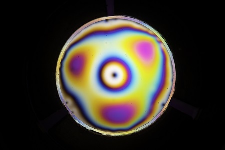
The spatially varying birefringence phase plate has a birefringence distribution with trigonal symmetry. In effect, it can produce beams that have every possible polarization state. Courtesy of J. Adam Fenster/University of Rochester.
CHIDO features a glass plate subjected to uniform physical stress all around its periphery. The device is placed in the Fourier plane at the back of a fluorescence microscope, where it is able to transform the image of a single molecule into a distorted focal spot. The shape of the molecule directly encodes the 3D information.
The microscope, with CHIDO incorporated, is then able to precisely monitor the position, orientation, and oscillations of single cells.
When a protein changes shape, it exposes other atoms that enhance the biological process, so the change of shape of a protein has a significant effect on the other processes inside the cell, said Brasselet, director of the Fresnel Institute. A way to monitor this change of shape is to use CHIDO to observe the orientation of fluorescent molecules attached to the protein under investigation.
The spatially varying birefringence phase plate has a birefringence distribution with trigonal symmetry; it can produce beams that have every possible polarization state.
“This is one of the beauties of optics,” Brown said. “If you have a device that can create just about any polarization state, then you also have a device that can analyze just about any polarization state.”
The origin of the device stems from an investigation of unusual polarization patterns in optical beams, some of which exhibited a spoke-like radial pattern. Ph.D. student Kathleen Youngworth demonstrated on a tabletop that, when tightly focused, the beams exhibited polarization components that pointed in almost any direction in three dimensions.
Another Ph.D. candidate, Alexis (Spillman) Vogt, attempted to replicate the effect by applying physical stress to the edges of a glass cylinder.
“Metal expands at a faster rate when you heat it than glass does,” Brown said, “and so you could heat the glass and metal up very hot, insert the glass in the middle of the metal, and as it cools down, the metal would shrink and create a tremendous force on the periphery of the glass.”
The group then fabricated a selection of its samples, fitting them in metal rings so that they could be used with a confocal microscope, which involved heating the components.
In the process, the group inadvertently applied more stress than called for with one of the plates, and, as soon as a team member handled it, the plate exhibited unusual and distinct qualities. The Rochester researchers introduced the term “stress engineered optic” to describe the elements. Later, they realized that the windows could be used to explore additional and even untested concepts in microscopy.
The research was published in Nature Communications (www.doi.org/10.1038/s41467-020-19064-6).