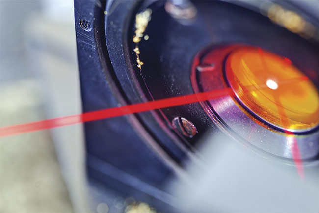
Fiber lasers provide high resolution, high contrast, and deep penetration for applications that aid in medical evaluation.
AMIRA TANDIROVIC GURSEL, ADANA ALPARSLAN TÜRKES SCIENCE AND TECHNOLOGY UNIVERSITY
According to the latest reports from the United Nations Department of Economic and Social Affairs and the World Health Organization (WHO)1,2, life expectancy has more than doubled in the last two centuries, from less than 30 years to over 70, thanks to both life-saving scientific innovations and health improvements. But the rise in life expectancy increases issues of morbidity, primarily driven by the high prevalence of chronic diseases, such as cardiovascular disease, cancer, diabetes, and arthritis. This has serious consequences for nations’ health and health care systems.
Reducing the incidence of chronic disease and further improving survival rates are not possible without timely and accurate diagnoses. A critical starting point involves detecting, locating, and accurately evaluating disease in its initial stages — in organs and tissue, down to cellular or even molecular levels — before the onset of symptoms.
One important step in tackling these challenges will be developing and advancing light-based technologies — such as near-infrared (NIR) and mid-infrared (MIR) spectroscopy, and NIR and MIR microscopy — for high-precision molecular sensing. These technologies could provide complete information on the chemical composition of biological samples to aid in early-cancer diagnostics, biofluid screening, breath analysis, and inspection of bodily fluids. In this context, the fiber laser is currently in the spotlight as an outstanding detection tool.
The first working fiber laser was developed by Elias Snitzer and colleagues in the 1960s as a result of the brilliant idea to combine two then-young technologies: fiber and the laser. The fiber laser wasn’t proved to be useful, however, until around 1985, when it was rediscovered by David Payne and colleagues at the University of Southampton in England. Although originally intended for telecommunications applications, fiber lasers caught the research community’s attention in short order.
Since then, thanks to important advantages — such as compactness and simplicity; excellent beam quality; high efficiency; and widely adjustable output power, pulse duration, and repetition rate — fiber laser technology has been pursued vigorously and is thus becoming more common.
In spectroscopy, where high tunability is required, fiber lasers are considered an obvious alternative to conventional light sources — particularly quantum cascade lasers (QCLs). Femtosecond lasers, with their spectral coverage no longer limited by the use of silica fibers, could become ideal fiber laser sources of supercontinuum and optical frequency combs. It should be noted that fiber lasers’ wavelength tunability could be additionally improved through the laser system design.
Theoretical background
Fiber lasers are a class of solid-state lasers that uses a rare-earth-doped core of optical fiber as an active gain medium. An optical fiber becomes active by doping its core with one or more rare-earth ions. Some ions of the host medium are replaced by the trivalent rare-earth ions of the same valence and similar size, to reduce the influence of the host lattice on the wavelengths, bandwidths, and cross sections of the relevant optical transitions3. Table 1 shows a list of rare-earth elements and common host media, as well as important emission wavelengths in the NIR and MIR regions.
TABLE 1
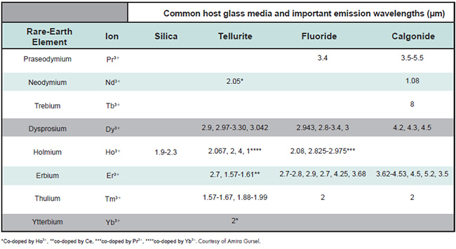
Figure 1 shows an example of a cladding-pumped Fabry-Pérot laser with fiber Bragg gratings (FBGs) and fiber configuration, along with a schematic of the double-clad structure and its cross section and transverse refractive index profile on the upper right corner of the figure. The FBGs are used as both the feedback device — in place of conventional dielectric mirrors — and an output coupler.

Figure 1. A schematic of a Fabry-Pérot cavity with fiber Bragg gratings and double-clad fiber, showing the end view and index profile. Courtesy of Amira Gursel.
Fiber lasers mainly use double-clad fiber designs consisting of three layers — core, inner cladding, and outer cladding — made of materials with various refractive indices to form a waveguide. The propagation of pump light is restricted to the single-mode core, where it can be absorbed by the laser-active ions, and the inner cladding is restricted by an outer cladding with a lower refractive index. A significantly larger area, as well as a much higher numerical aperture of the inner cladding, allows the more efficient launch of the output, which in turn provides the high beam quality that is strongly desirable for medical applications. Fiber lasers are constructed monolithically; that is, all of the fiber components are spliced together, resulting in more robust and longer-lasting laser tools compared to their counterparts, such as doped insulator lasers, diode lasers, QCLs, and interband cascade lasers (ICLs).
NIR to MIR
The use of the NIR regions, spanning between 800 and 2500 nm, has been widely implemented in nondestructive analysis of bulk materials. The medical community has noticed the lasers’ low cost, simplicity, safety, and high signal-to-noise ratio (all thanks primarily to recent advancements in microelectronic components) combined with the fact that transparency of tissues reaches its maximum in the NIR. Fiber lasers are thought to be the most promising candidate for noninvasive monitoring of physiological parameters, including glucose.
NIR spectroscopy is a typical vibrational spectroscopic method that uses overtone or harmonic vibrations, which are associated with a change in the dipole moment of the molecules. With regard to skin, light within the NIR region has a penetration depth inversely proportional to the wavelength, ranging between 1 and 100 mm, a range that is enough to reach the skin’s vascular region.
Fiber lasers in the NIR region, already used in a vast array of important applications, could also be used in transformative applications in the MIR region, for instance, as the light source for breath analysis and precision spectroscopy.
MIR spectroscopy involves quantification of the composition of the sample measured by light ranging between 2.5 and 25 µm of the electromagnetic spectrum. Broad spectral coverage of the MIR region provides access to chemical “fingerprints” of many molecules, which can be recognized with great sensitivity. MIR spectroscopy can also be used to identify even tiny quantities of chemicals associated with many diseases.
Until a few years ago, poor coherence of light sources and troublesome sensing due to highly attenuated backscattered signals kept MIR spectroscopy out of wide use in diagnosis. However, in recent years, new developments in lasers and sensing techniques have brought about significant improvements. MIR photonic components have become diversified, miniaturized, and integrated, paving the way for enhancement of the more efficient light sources.
In this context, it should be emphasized that the project MINERVA (MId- to NEaR infrared spectroscopy for improVed medical diAgnostics), funded by the European Commission, brought MIR spectroscopy back into the limelight by opening up the MIR region for rapid medical imaging. One of the primary driving forces of the project was the lack of practical light sources, components, detectors, and modulators to access the MIR region. Thirteen project partners from across Europe came together with a common objective: to develop photonic technology that improves the biomedical imaging required for early cancer diagnosis, which is often key to survival4.
The fruit of this teamwork is an imaging system — the first demonstration of high-resolution, multispectral supercontinuum spectroscopic tissue imaging across the MIR — that offers two specific high-impact applications: high-volume pathology and noninvasive in vivo skin cancer screening. The results of the project are important not only for diagnostics but also the scientific community in general. In terms of the application of laser fiber technology in diagnostics, the project appears to be an important step forward. According to its final report, fiber lasers were usually used as the pump sources for ultralong-wavelength supercontinuum generation. One was a 2.9-µm Q-switched Er:ZBLAN, with 4.5-µm pulses, developed by LISA Laser with components from G&H, used as a primary MINERVA pump source for the 3- to 9-µm supercontinuum. The other was a mode-locked PR-doped chalcogenide fiber laser pumped by a 2-µm Tm-doped fiber laser, used as a primary MINERVA pump source for the 3- to 12-µm supercontinuum and developed by DTU Fotonik and the University of Nottingham5.
Photoacoustic microscopy
Photoacoustic microscopy (PAM) combines optical and ultrasound imaging,
providing ultrasonic spatial resolution and high optical contrast. Its hybrid nature may help it to gain a competitive edge over other modalities. When tissue is illuminated by a short laser pulse, the absorbed light leads to a thermoelastic expansion, which in turn converts to an acoustic emission, generally in the megahertz range. The resulting ultrasonic waves are detected conventionally by ultrasonic transducers.
Recorded signals can be used to map the distribution of the precise locations of the optical absorbers. Relatively low scattering of ultrasonic waves in biological tissues provides deeper imaging penetration beyond the optical transport mean-free path. The contrast of PAM is internally produced by the optical absorption of chromophores within the tissue.
The ideal PAM light source produces short enough pulses (i.e., several nanoseconds) for generating photoacoustic signals efficiently, and it emits wavelengths in both the visible and NIR regions, from 600 to 1200 nm. The visible range covers absorption peaks of tissue chromophores in their spectra, while the NIR region is required to obtain adequate penetration depth. The repetition rate of the pulses generating the light source should also be in at least the kilohertz region to obtain fast imaging.
The light source should be widely tunable to provide analysis of as many types of substances as possible. For example, a laser suitable for melanoma cell detection should also be appropriate for hemoglobin imaging. The light sources used in PAM are typically Q-switched lasers, low-
power laser diodes, and, recently, fiber lasers. But none of these offers independently adjustable key parameters.
To overcome this problem, the BUMILAB research group at Bogaziçi University in Istanbul developed a fiber laser with independently adjustable properties6. A tunable fiber-based MOPA laser system (Figure 2) produces nanosecond pulses, covering the spectrum from 450 to 1100 nm, specifically for PAM. The supercontinuum part of the system is all-fiber-integrated; guided beam propagation renders it misalignment-free and largely immune to mechanical perturbations. Free-space harmonic generation creates higher pulse energy for a specific band — that is, 532 nm — and also generates
UV light with wavelengths of 355 and 266 nm. The tunability of the laser parameters enables the use of only one laser for many various PAM applications, with a high repetition rate for fast scanning.
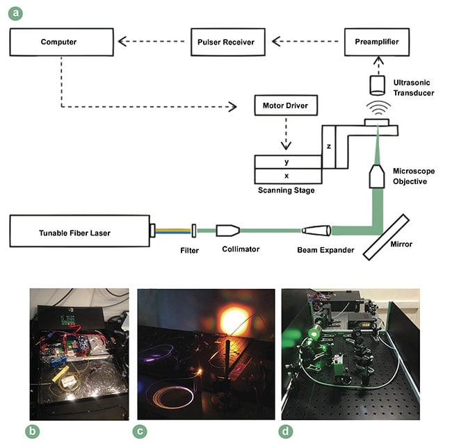
Figure 2. The setup of an optical resolution PAM system that uses a tunable fiber laser (a), a compact fiber laser system for PAM (b) with the outputs of:
supercontinuum (c) and harmonic generation units (d). Adapted with permission from Reference 6.
Breath analysis
Breath analysis is an application for quick noninvasive metabolic monitoring that can be used in disease diagnostics. It can assist in tracking disease progression and monitoring therapeutic interventions.
Although the importance of the exhaled human breath was recognized by Hippocrates, who realized that some diseases have a characteristic odor, it was kept from wide use in diagnosis until the end of the 20th century, primarily due to the poor sensitivity of many traditional analytical methods. However, intensive research on the origins, pathways, and pathophysiological roles of the breath components; advancements in optical methods using NIR and MIR sources; and a significant improvement in gas-sensing performance have added new dimension to breath analysis. These developments are leading the way toward real-time, user-friendly point-of-care diagnosis.
Exhaled human breath contains up to 3500 compounds, including exogenous, inorganic, and volatile organic compounds. Some of these are important biomarkers that may be used alone or in combination with others to assess the state of a patient’s health. With each exhalation, thousands of biomarkers are ejected with the breath, providing an individual “breathprint” that reveals even minimal change in the internal metabolism as result of illness, as well as the response to various external stimuli, including any change in the physical or chemical structure of an organism’s external environment. Almost 40 biomarkers — including acetone, ethanol, carbonyl sulfide, ammonia, and nitric oxide — have NIR or MIR spectral fingerprints that can be successfully analyzed7.
Breath analysis that is acceptable for diagnostics requires high sensitivity and
fast response time, and just a few spectroscopic techniques are able to meet these demands. Breath analysis applications require broad wavelength tenability from light sources. Although diode lasers are still the conventional light source for exhaled breath analysis, ICLs and QCLs are gradually giving way to fiber lasers. Figure 3a shows a fiber laser as a swept-wavelength source for ammonia gas sensing, and Figure 3b shows a fiber laser in supercontinuum-generation stage8,9.
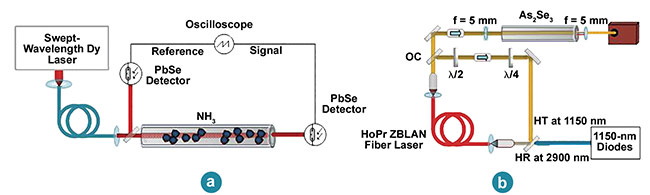
Figure 3. A swept-wavelength Dy:ZBLAN fiber laser source for sensing of ammonia gas (a) and a Ho3+, Pr3+-co-doped ZBLAN fiber laser (b) in
supercontinuum-generation stage. Adapted with permission from References 8 and 9.
Other fiber lasers used to analyze breath are NIR tunable Er3+-doped fiber lasers in combination with optical fiber amplifiers; broadband Tm/Ho-doped fiber lasers tunable from 1.8 to 2.09 µm; ultrafast Dy3+-doped fiber lasers beyond 3 μm; and Tm-doped fiber lasers that are ideal for mid-IR supercontinuum generation9,10.
Features such as straightforward and efficient optical excitation, compactness,
robustness, relatively easy heat management, and especially wavelength tunability of fiber lasers exhibit clear advantages over other laser technology. However, such rapid and accelerating acceptance of fiber lasers seen in fields as diverse as materials processing, telecommunications, and defense-related applications has not been seen in the medical community.
When it comes to the MIR light sources that are strongly required for diagnosis, up to now, advancements have been predominantly associated with the development of QCLs, which currently form the basis of MIR photonics.
But QCLs are too bulky and less efficient due to producing a large amount of heat, whereas fiber lasers show quite satisfactory results. While the value of fiber lasers has been widely recognized in scientific research, they are not yet commercially available at the desired level, contrary to expectations, primarily because of the high cost of fiber, especially glass fiber in the MIR region. Fortunately, significant research directed toward the development of novel glasses, as well as the improvement of existing ones, will doubtless have a positive impact, not only on moving further into the MIR region but also on diversifying fiber lasers, which in turn could make their cost come down.
It would not be surprising if fiber lasers
were soon used as next-generation coherent light sources for new spectroscopy and sensing technologies in many diagnostic applications.
Meet the author
Amira Tandirovic Gursel, Ph.D., is an assistant professor at Adana Alparslan Türkes Science and Technology University. She received her doctorate in 2015 from Gaziantep University and works in medical electronics and photonics.
Acknowledgments
The author thanks Seydi Yavas as well as the BUMILAB research group for sharing their results and for their valuable contributions to the work presented here.
References
1. United Nations (2019). 2019 Revision of World Population Prospects, https://popula
tion.un.org/wpp.
2. World Health Organization (January 2017). World report on ageing and health. Indian J Med Res, Vol. 145, Issue 1, pp. 150-151.
3. A.T. Gursel (July 25, 2018). Fiber lasers and their medical applications. In Optical Amplifiers — A Few Different Dimensions, www.doi.org/10.5772/intechopen.76610.
4. MINERVA (September 2017). MINERVA project newsletter #8. www.minerva-project.eu/wp-content/uploads/2017/10/MINERVA-newsletter-8.pdf.
5. A.B. Seddon (March 4, 2019). Progress in biomedical mid-infrared hyperspectral imaging with fiber-based supercontinuum laser light. Proc SPIE, Vol. 10873. In Optical Biopsy XVII: Toward Real-Time Spectroscopic Imaging and Diagnosis.
6. E. Aytac-Kipergil et al. (2016). Development of a fiber laser with independently adjustable properties for optical resolution photoacoustic microscopy. Sci Rep, Vol. 6, article 38674.
7. B. Henderson et al. (2018). Laser spectroscopy for breath analysis: towards clinical implementation. Appl Phy B, Vol. 124,
No. 8, p. 161.
8. R.I. Woodward et al. (2019). Swept-wavelength mid-infrared fiber laser for real-time ammonia gas sensing. APL Photonics, Vol. 4, Issue 2, 020801.
9. D.D. Hudson et al. (2017). Toward all-fiber supercontinuum spanning the mid-infrared. Optica, Vol. 4, Issue 10, pp. 1163-1166.
10. R.I. Woodward et al. (March 2019). Mid-infrared fiber sources for real-time biomedical sensing. Proc SPIE, Vol. 10873. In Optical Biopsy XVII: Toward Real-Time Spectroscopic Imaging and Diagnosis.