New developments in imaging technology, in combination with advanced microscopy techniques, are enabling new applications for fluorescence sensing.
JASPER BOSCH, LAMBERT INSTRUMENTS BV
Detecting fluorescence light via imaging and fluorescence sensing can take researchers and clinicians into the body, allowing them to visualize samples and biological progression of areas such as cancer detection or cardiovascular function. The fluorescence light contains the information about the sample that a fluorophore has detected, so recording it with great precision is important for the accuracy of the measurement.
In recent years, there have been many improvements in the fluorophores and imaging technology involved in fluorescence sensing. These developments have improved the capabilities of fluorescence detection and are enabling new in vivo imaging applications. For example, by combining light sheet fluorescence microscopy with super-slow-motion imaging, it is now possible to construct a 3D representation of a sample. And improvements in fluorescence lifetime imaging are enabling researchers to study molecular interactions and physicochemical parameters, such as oxygen concentration or pH, with much better resolution and shorter time intervals.
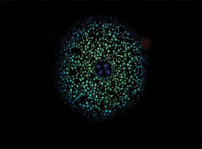
Stitched fluorescence lifetime image of a ranunculus sample recorded with the Lambert Instruments FLIM Attachment (LIFA). The fluorescence lifetime is presented as a pseudo-color overlay on the image, with shorter lifetimes shown as blue and longer lifetimes shown as green. Courtesy of Lambert Instruments.
Imaging with fluorescence light requires optical instrumentation to isolate it from excitation, autofluorescence, and background light. This is usually done with optical filters that transmit light only within the wavelength range of interest. An imaging device captures the transmitted light, and the image is presented to the user on a computer display. This image represents the information that the fluorophore communicates through fluorescence light.
Such light is often quite dim, as its intensity is limited by factors such as the fluorophore concentration, quantum yield, molecular state, and excitation light level. This dim light is recorded by an imaging device — one that is able to record images in low-light situations. However, any image unavoidably contains some noise as a result of imperfect optical filters, unwanted reflections, or image sensor noise.
The quality of the image depends on how well the fluorescence light can be distinguished from the noise. This ability is quantified by the signal-to-noise ratio (SNR) — the greater the SNR, the better the image quality. To optimize this setup, both the fluorescence emission and detection should be considered. Increasing the excitation light will increase the emission light as well. However, there is a limit to the excitation light levels a biological sample can endure. Beyond a certain level, the effects of photobleaching and photodamage may reduce the brightness of the fluorescence emission light and subsequently the quality of the recorded image.
The properties of the fluorophore also should be considered. In recent years, fluorophores with higher quantum yield have been developed1,2. Such fluorophores convert excitation light into emission light more efficiently and therefore emit more fluorescence light than other fluorophores upon excitation with the same light levels. This increases the fluorescence light output of the sample without any detrimental effects.
On the image detection side, the optics and optical filters that form the light path should be optimized to ensure that as little light as possible is lost in transmission from the sample to the imaging device. Figure 1 demonstrates an imaging system for fluorescence sensing where a single focal plane of a sample is being imaged with a confocal spinning-disk microscopy setup.
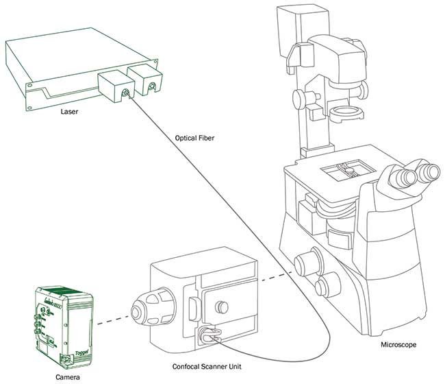
Figure 1. Schematic overview of an imaging setup for fluorescence sensing. Excitation light from a laser is transmitted through an optical fiber to a confocal scanner unit. The excitation light passes through a Nipkow disk and into a microscope. Fluorescence light from the sample is isolated from the excitation light by a dichroic filter in the confocal scanner unit. The Nipkow disk blocks out-of-focus light and transmits only light from a single focal plane to the camera. Courtesy of Lambert Instruments.
New developments in image sensor technology have improved the light detection efficiency of imaging devices. Scientific CMOS (sCMOS) and electron-multiplying CCD (EMCCD) sensors exhibit low noise levels and can record clear images in low-light conditions because of their low SNR. A common way to further reduce the image sensor noise is by cooling the image sensor. This reduces the probability of spontaneous generation of charges in the semiconductor material of the image sensor known as dark current. Minimizing such noise sources improves the SNR of the recorded images.
Slow-motion fluorescence
Improved light sensitivity of image sensors not only improves the quality of the recorded images, it also enables imaging at higher frame rates to improve the temporal resolution of the recorded image data. Current sCMOS and EMCCD sensors capture images at up to 100 fps, but CMOS sensors can reach frame rates of thousands of frames per second. These sensors exhibit higher noise levels than sCMOS and EMCCD sensors, although ongoing development has improved their light sensitivity. As a result, these image sensors now require less time to gather an equivalent amount of light as older sensors in the same light conditions. Therefore, images of the same quality can be captured at higher frame rates.
Slow-motion imaging technology has seen great improvements over the past decade as well. Now, even smartphone cameras record video at rates of hundreds of frames per second. High-end cameras can capture super-slow-motion video at tens of thousands of frames per second. The limiting factor in such cameras had been the size of the internal recording memory, which would fill up rather quickly because of the large amount of image data recorded per second. Now, however, with the increasing throughput speed of solid-state drives, the image data can be written directly to a drive rather than recorded first to random-access memory. This increases the ease of use and extends the possible duration of super-slow-motion recordings from fractions of seconds to several minutes.
Super-slow-motion cameras can be combined with analog light amplification technology, such as an image intensifier, to boost the light levels before they are detected by an image sensor. With the increased light sensitivity of image intensifier technology, it is now possible to record fluorescence images at frame rates of thousands — or even tens of thousands — of frames per second. The Lambert Instruments HiCAM Fluo shows that this combination of technologies can enable applications such as in vivo super-slow-motion imaging and detailed 3D imaging with fluorescence light.
In vivo super-slow-motion imaging provides a new way to look at biological samples. By slowing down the playback speed of a video, it is now possible to see what happens during fast dynamic processes. As shown in Figure 2, the red blood cells of a zebrafish were labeled with the DsRed fluorophore to study its cardiovascular system (bottom right corner of images), and were imaged with an intensified high-speed camera at 2000 fps. In Figures 2a-d, red blood cells are transported from one chamber to the next and into the aorta, which is shown in Figure 2e. This methodology provides an interesting new visualization for the study of cardiovascular disease.
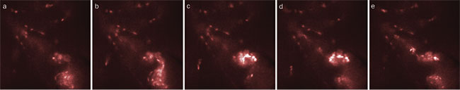
Figure 2. Red blood cells in the cardiovascular system of a zebrafish are labeled with the DsRed fluorophore and imaged with an intensified high-speed camera. Red blood cells are transported from one chamber to the next (a-d) and into the aorta (e). These images were recorded with an interval of 25 ms between subsequent images and with an exposure time of 500 µs to prevent motion blur. Courtesy of Max Planck Institute for Heart and Lung Research.
The same methodology can be applied in imaging electric signals in the brain or the heart by using a voltage-sensitive fluorophore. A super-slow-motion recording of a sample with such a fluorophore can show how a voltage change propagates through the sample.
Another field of research where super-slow-motion imaging can provide new insights is microfluidics for lab-on-chip applications. Because of the size of the microchannels, the number of light-emitting particles is low, as is the corresponding fluorescence light level. However, the particles do move relatively fast. To properly record the emitted light with minimal motion blur, a fast camera with extreme light sensitivity is essential.
Fast FLIM
Other developments in image sensor technology include the ability to implement advanced detection methods in image sensors. An example of this is the development of new image sensor technology for fluorescence lifetime imaging microscopy (FLIM).
The fluorescence lifetime is the quantity that describes the rate of decay of the fluorescence light intensity after a fluorophore is illuminated with excitation light. Many specialized fluorophores exist that encode environmental parameters, such as oxygen concentration or pH levels, in the lifetime of the fluorescence light they emit.
FLIM camera technology for wide-field imaging previously required multiple devices for signal generation and demodulation of the detected fluorescence light. Next-generation image sensor technology makes it possible to implement the demodulation in the image sensor itself. This not only reduces the size of a FLIM setup and associated costs, it also increases photon efficiency and the image acquisition speed.
Such enhancement of imaging and sensing instrumentation from companies such as Lambert Instruments can record quantitative fluorescence lifetime images at higher frame rates. With this improved temporal resolution, new applications of FLIM are now being explored in live-cell imaging3 and single bacterial cell imaging4. Fluorescence lifetime imaging at these frame rates assists researchers in unveiling the characteristics of dynamic processes in living cells in more detail.
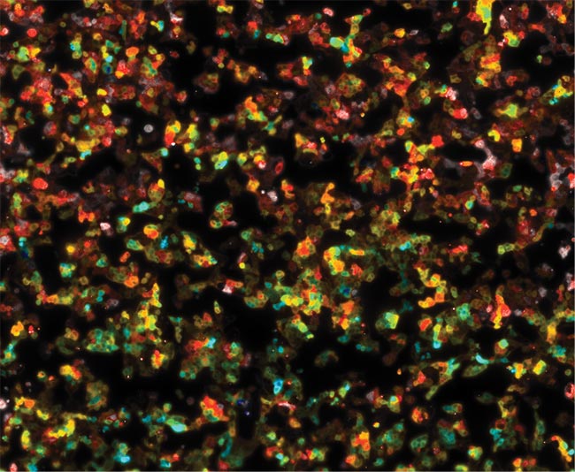
Figure 3. Stitched fluorescence lifetime image of a population of HAP1 cells that express an EPAC-sensor, which shows an increase in fluorescence lifetime when the concentration of cyclic-AMP increases. The expression level varies from cell to cell. The fluorescence lifetime is presented as a pseudo-color overlay on the image, with shorter lifetimes shown as blue and longer lifetimes shown as red. Courtesy of the Netherlands Cancer Institute.
Fast fluorescence lifetime imaging, in combination with a motorized microscope stage, makes it easy to quickly image large samples or multiwell plates (Figure 3). This enables researchers to gather more empirical data to achieve statistically better results.
Meet the author
Jasper Bosch is a development engineer at Lambert Instruments BV. He focuses on research and development for the company’s fluorescence lifetime imaging microscopy and intensified high-speed imaging products; email: [email protected].
References
1. D. Bindels et al. (2017). mScarlet: a bright monomeric red fluorescent protein for cellular imaging. Nat Methods, Vol. 14, pp. 53-56.
2. J. Goedhart et al. (2012). Structure-guided evolution of cyan fluorescent proteins toward a quantum yield of 93 percent. Nat Commun, Vol. 3.
3. M. Raspe et al. (2016). siFLIM: single-image frequency domain FLIM provides fast and photon-efficient lifetime data. Nat Methods, Vol. 13, pp. 501-504.
4. J. Detert Oude Weme et al. (2015). Single cell FRET analysis for the identification of optimal FRET-pairs in Bacillus subtilis using a prototype MEM-FLIM system. PLOS ONE, Vol. 10, Issue 4.