Brent D. Johnson, Senior News Editor
Some researchers are legendary for their lab capability, easily able to find needles in haystacks. Others who complain that they were never good in the lab appreciate simple-to-operate equipment. Although the concept of point-and-shoot microscopy may offend the talents of the inveterate microscopist, recent advances in automation and ergonomics will likely win the hearts of fledgling experimenters and veterans alike.
In May, Leica Microsystems Inc. of Bannockburn, Ill., introduced a digital microscope series that targets both biological applications (models DM4000 B and DM5000 B) and materials science (DM4000 M). In each case, the microscope, camera, software and image database are available as a complete package.
Common to these systems are features such as automated aperture, light adjustment and contrast methods; color-neutral brightness control; and condensers, all of which facilitate configuration of optimal settings. It also is possible to set up complex procedures automatically, so researchers don’t have to break their concentration to focus on operating the instrument.
A fluorescence intensity manager sets the level of fluorescence illumination required in various life sciences applications. Software also controls color neutralness and eliminates the need for time-consuming Köhler illumination adjustment of the microscope. In addition, it is now possible to automatically adjust the system to always work with the objective and magnification commonly selected by each user.
Spectral confocal microscopy
Another recent development is a confocal microscope that replaces conventional beamsplitters with a programmable device (the TCS SP2 acousto-optical beamsplitter). Beamsplitters reflect exiting light toward a sample and transmit the emitted fluorescence for detection. Programming capability allows fast, precision adjustment from single to multiple chromatic characteristics of any type. This enables operating the device across the entire visible wavelength without filters.
The system resembles an infinite set of multichroic beamsplitters and is designed as a spectral imaging and measurement environment for multifluorescent samples. In comparison with conventional beamsplitter-equipped confocals, it facilitates automatic adjustment for selected laser lines, rapid wavelength selection and greater transparency. When combined with an emission side prism spectrophotometer and a selectable emission window, the microscope gives the user the freedom to combine dyes for confocal analysis.
Forensic evaluation
Another area of microscopy gaining prominence involves forensics. This application is unique in that the samples observed are not merely passive specimens. Instead, they have the potential to incriminate or exonerate. A synthetic fiber, a drop of blood or a bullet fragment, properly identified, can mean the difference between corroborating the story of an innocent bystander and convicting a felon. Under these circumstances, the microscopy technique must be nondestructive to preserve the evidence. In addition, the image results must be sharp and recognizable to laymen.
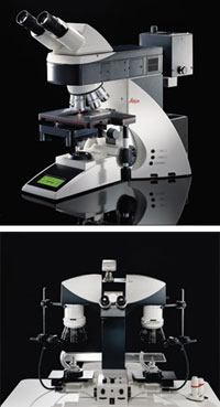
Leica forensic tools are automated to enable extremely precise firearm and toolmark examination.
Comparison microscopes and macroscopes have been available for firearm and toolmark examination for more than 90 years, but they have received a recent performance boost from automation. An example is a macroscope that has telecentric Plan Apo chromatically corrected objectives with fixed magnifications. Each objective has its own iris diaphragm. In addition, a built-in 1.5× magnification changer expands the range from 4× to 60× and provides up to eight discrete settings to ensure measurement reproducibility of fine details such as striations in fired bullets. The user calibrates the system once, and the exact objective measurement is maintained to ensure that conditions are identical whenever a sample must be examined.
An intelligent automation feature allows adjusting the speed of X-, Y- and Z-axes based on the objective magnification in use. The control software also synchronizes stage movement, control-lamp intensity, comparison bridge positioning and magnification. A single button switches from the superimposed image to side-by-side examination of questioned documents or split-image comparison of bullet cartridge caseor firing pin impression, and a multifocus module calculates a two-dimensional picture from several images.
Contact: Pam Jandura, Communications Department, Leica Microsystems Inc., Bannockburn, Ill.; +1 (847) 405-7062; fax: +1 (847) 405-0164; e-mail: [email protected].