As criminal cases pile up, automation and digital imaging techniques will help crime scene investigators close them more quickly.
Forensic crime shows such as CSI have taken over TV networks worldwide, providing viewers with a stylized glimpse into forensic science. A vital part of an investigator’s armory is the forensic microscope, crucial for inspecting trace evidence and for making bullet and tool mark identifications. For a century now in forensic biology, light microscopy has been used to identify spermatozoa in sexual assault cases.
Sadly, real-life crime laboratories often have hundreds of thousands of cases in vaults that have not yet been opened – some laboratories can even be years behind, said Richard E. “Dick” Bisbing, a forensic scientist for more than 40 years and formerly with the Michigan State Police. He is now executive vice president of McCrone Associates Inc. and an instructor at Hooke College of Applied Sciences LLC, both in Westmont, Ill.
“There are horror stories about criminals roaming free because there are inadequate resources and too many samples unworked,” he said.
Digital imaging
Consequently, forensic microscopy makers are looking to advance the development of more highly automated systems so that laboratory technicians can process samples more quickly, easily, accurately and less expensively.
Recent advances include combining optical work with spectroscopy and spectrophotometry for identifying fibers, paints and polymers, but the biggest change has occurred in the past few years: digital imaging.
Digital imaging via instruments such as the new Olympus polarized light microscopes BXiS and BX53 means that microscopic images can be captured with the touch of a button, then automatically labeled and archived together with related sample and analytical information.
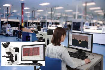
Tools such as the Olympus BXiS microscope, when used with Stream software, provide a comprehensive instrument for analysis of forensic samples. A cross section of a composite material is examined and measured using the Olympus BX51M microscope and Stream software. Courtesy of Olympus America Inc.
“Back when we had to capture images on film, documentation was often lacking. Today, most laboratories take photos of everything because they have high-quality imaging systems built onto their microscopes,” Bisbing said. “That’s been a big change in forensic science in just the past four or five years.”
Thanks to the microscope’s coded nosepiece, all the calculations and magnifications are input automatically.
“What this means in the laboratory where imaging is done is a much lower cost per image, along with the ability to store multiple images in multiple places so data is never lost,” said Matt Smith, director of sales and marketing, Scientific Equipment Group – Industrial, of Olympus America Inc. in Center Valley, Pa. “Hardware is only half a product now – users are really buying an integrated system.”
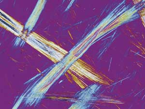
Combining optical techniques with infrared spectroscopy allows scientists to identify paints, adhesive tapes, minerals and related samples. In addition, Raman spectroscopy techniques are growing in popularity.
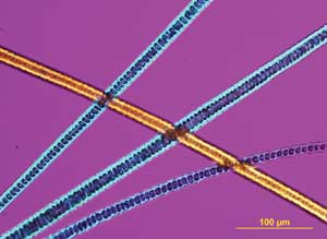
Asbestos fibers are typical specimens for forensic microscopes. Courtesy of Olympus America Inc.
In some cases, forensic scientists use a light microscope to make initial comparisons and select an appropriate portion of the sample for analysis, and then use another technique for some of the more detailed work.
The integration of spectroscopy with microscopes is expected to become more refined, as is the streamlined integration of entire systems, including software, hardware and storage.
“The more efficient and less costly computer power becomes, the faster this change will occur,” Smith said. “In addition, creating databases that can be queried easily is an area of importance. Because photography increasingly is a part of the work flow, digital camera technology will continue to develop, with cameras like the Olympus DP26, DP72 and other cameras that are more accurate, provide better color rendition and are push-button easy.”
Chromosome sequencing advances
On the biological side, there have been advances in chromosome sequencing, and there is progress in performing intracellular work with light microscopy. Fluorescence and other light microscopy techniques can now be applied to smaller samples, down to individual cells.
From the toolmakers’ and manufacturers’ perspectives, one of the greatest changes is that the spectrographic component, the optical components and all the resulting data now can live in the same system using the same database, along with images.
“This is powerful because all the techniques can be integrated on a single platform such as the BXiS microscope, used with Stream software, to speed up the work flow for forensic scientists,” Smith said. “Olympus also partners on an OEM basis with some excellent FTIR [Fourier transform infrared] and Raman systems, and all the data from all of these techniques lives in a single database.”
A microscope magnifies cat hairs. Courtesy of McCrone Microscopes & Accessories.
Polarized light microscopes and other modular instruments on the market accept polarized light accessories but also can be used for phase contrast, fluorescence and other techniques used by biomedical researchers and forensic biologists, according to Jeffrey McGinn, president and director of instrument sales at McCrone Microscopes & Accessories Inc. in Westmont, a sister company of McCrone Associates.
“Comparison microscopes and stereomicroscopes also are used in forensic applications,” McGinn said. “Additionally, stereomicroscopes are always sold with the polarized light systems. The stereos are used for sample examination with a larger field of view and the true 3-D perspective offered by stereoscopic viewing.”
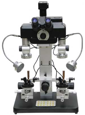
Unitron’s comparison forensic microscope was designed with the help of police organizations, forensic scientists and educators in the field of forensic science to inspect ballistics and firearms and to make tool mark examinations. Courtesy of Unitron Ltd.
Unitron Ltd. of Commack, N.Y., recently launched a comparison forensic microscope (CFM) and is working on approvals to place it into governmental facilities. The CFM was designed with the help of police organizations, forensic scientists and educators in the field of forensic science to inspect ballistics and firearms and to make tool mark examinations.
Evidence can be observed simultaneously via the comparison bridge that supports two sets of matched objective steps on a five-step magnification changer. The microscope series, equipped with 22-mm field-of-view eyepieces, produces erect, unreversed images that move in the same direction as the specimen for ease of use. The images can be viewed at 100 percent right or left side, split screen or superimposed.
“I took our CFM to a forensic conference a few months ago and received nothing but kudos from the state police, sheriff’s department and local Bureau of Investigation,” said Heston Singh, a business developer at Unitron. “Our products are considered mechanically and optically competitive … and are considered far superior to most of the products coming out of the Pacific Rim.”
Beyond CSI
It’s not just crime scene units that make use of forensics: The FBI and other federal entities, private firms, private laboratories and even industrial laboratories use forensic instruments. Some industrial forensic work lies in research and development, and some is used to solve problems relating to litigation and safety because some events can be understood only by studying a material closely and identifying its origin or composition.
Hooke College and McCrone Microscopes & Accessories are carrying out extensive US National Guard training, working with first responders to teach them essential techniques to identify samples and assure safety when working with unknown and potentially hazardous materials.
“When there is an incident, they show up on the scene and aid in homeland defense,” McGinn said. “We continue to train these teams, who come to our facility for basic and advanced microscopy courses.”
Other future developments could come from projects such as the cooperative agreement between the National Institute of Justice and Hooke College, in which 300 trace evidence examiners have been trained so far in the use of light and electron microscopes, along with Raman and infrared spectroscopy.
This is a development that can truly help advance the practice of forensic science, Bisbing said.