High-contrast imaging at the nanometer scale is increasingly critical for progress in a broad range of areas, including materials science, physics, biology, and chemistry.
WILL HARRIS, KMLABS INC.
Both optical and x-ray microscopy are well-established tools, but nanoscale imaging with light in the intermediate range — specifically the extreme UV (λ ~8 to 80 nm) — has been neglected. This has been primarily because of a gap in available tools and techniques, so thus far the potential for progress has been relatively untapped.
However, recent advancements in suitable lab-scale light sources, as well as in computational imaging approaches, such as lensless coherent diffraction imaging (CDI), now make it possible to measure structures and features at <15-nm resolution and with unique contrast sensitivity. These new sources and approaches also enable spectral and temporally resolved diffraction measurements that can elucidate even the fastest dynamics in materials.
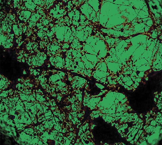
EUV ptychography reconstruction via CDI showing artefact-less reconstruction quality of the fine neural structures. Courtesy of Peter Baksh et al./
See Reference 3.
Imaging techniques at the microscale and nanoscale form an essential component of the experimentalist’s tool set for areas spanning from the biological to the physical sciences. Over time, a variety of techniques have been developed to address particular length scales, dimensions, and contrast modalities. Microscopy using visible light has both a long history and exciting new developments in wide-field, confocal, and (more recently) superresolution techniques. Concurrently, the evolution in electron microscopy over the past decades — from transmission (TEM), to scanning (SEM), and multibeam focused ion beam SEM (FIB-SEM) instruments — has pushed resolution to the atomic scale and offers an increasing number of contrast modalities. Furthermore, the increasing number of synchrotron facilities and lab-based x-ray tube sources has improved the accessibility of various x-ray techniques, mostly in the hard x-ray regime, including volumetric imaging measurements such as tomography.
For biological specimens, CDI in the EUV spectral range has been used for high-resolution imaging of fixed, embedded, and unstained neurons using transmission CDI.
In each case, the utility of the technique has been driven by the availability of sources of the given particle and wavelength, along with the necessity of imaging optics to properly manipulate the beam to form and detect an image.
Of notable absence to date from this array of imaging technologies is nanoimaging in the portion of the electromagnetic spectrum between visible wavelengths and x-rays — the spectral region spanning the vacuum UV (VUV) to extreme UV (EUV) and to the edge of the soft x-ray range (from approximately 8 to 250 nm, or equivalently, 5 to 150 eV).
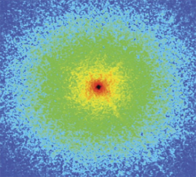
A typical individual diffraction pattern collected using a CDI approach with tabletop-generated EUV light. Such a pattern — representing a Fourier transform of the portion of the object illuminated in either transmission or reflection geometry — is directly acquired without any lens between the object and the detector. Courtesy of Kapteyn-Murnane Group/JILA, University of Colorado Boulder.
This gap in imaging capabilities is remarkable in that the wavelengths match the resolution requirements for many applications (resolution ≈ wavelength). This is also the range where electron-binding energies, bond dissociation energies, semiconductor bandgaps, and elemental absorption edges exist (including, for example, the lithium K-edge at about
55 eV), creating opportunities for producing image contrast for some of the fundamental building blocks of matter. This is particularly true for chemical structure contrast, and for functional characterization in localized measurements, especially for low-Z materials where electron microscopy often fails.
Unfortunately, this portion of the spectrum has historically been underexplored, with just a limited number of studies at synchrotron sources. Several reasons exist for this:
- Outside of a few select large-scale facilities, there have been few reliable light sources in this domain accessible to the general scientific community.
- While shorter or longer wavelengths (hard x-rays and visible light, respectively) are at least partially transmissive, photons in the VUV to soft x-ray range interact very strongly with most materials. This holds strong promise for identifying new contrast mechanisms, but also means the light is absorbed, which typically restricts signal to surface and near-surface regions. It also effectively impedes the design of lenses for this range.
- In addition to the sample itself, photons in this range are also strongly attenuated by air, so beams and measurements must be performed under vacuum or noble gas atmospheres, unlike with IR, visible, or hard x-ray.
The combination of these challenges has limited the reach of a multitude of experimental techniques. Photoelectron spectroscopy, including angle-resolved photoelectron spectroscopy (ARPES); different forms of absorption spectroscopy; and scattering, diffraction, reflection, and imaging methods have been applied at VUV/EUV synchrotron beamlines, but again, with limited access.
Recently, many of these challenges have begun to be overcome. The most notable advancement comes in the availability of suitable tabletop light sources. With the recent maturation and commercialization of laser sources at very short wavelengths (generated through nonlinear frequency upconversion, including high harmonic generation), robust VUV, EUV, and soft x-ray light are now available for the lab. These new sources are approaching turnkey reliability for use in a broad range of experiments without the need for prior expertise in laser physics or specialized optics.
For example, the XUUS high harmonic generation system from KMLabs Inc., together with a Ti:sapphire 800-nm pump laser of sufficient energy, produces highly coherent femtosecond-duration pulses of light with wavelengths that can span tens of nanometers. This opens the door to new spectroscopy and microscopy methods, including ultrafast measurements of time-resolved dynamic events on the femtosecond timescale using pump-probe techniques.
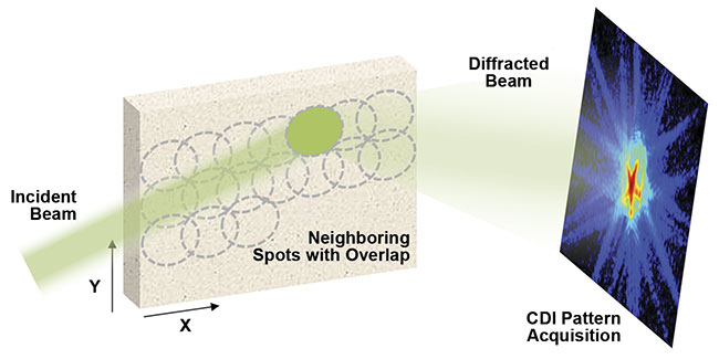
Acquisition scheme for CDI ptychography in a reflection geometry. A relatively large coherent laser
spot is rastered across the surface of the sample with overlap of neighboring spots. Ptychographic
reconstruction produces spatially resolved information about the amplitude and phase of the reflected wave, with spatial resolution that can exceed the wavelength if the detector captures a large scatter
angle. Courtesy of Kapteyn-Murnane Group/JILA, University of Colorado Boulder.
While necessity of a vacuum environment endures as a constraint for such experiments, intelligent engineering can maximize the flexibility and utilization of tabletop light-source instruments by embracing a flexible multibeamline/experiment concept. This concept is analogous to the model employed at synchrotrons with numerous beamlines and even multiple experimental hutches sharing a common beamline, or similarly, to electron microscopy where various optional detectors and modalities are available for different types of measurements. In a similar fashion to the synchrotron but on a much smaller (and more affordable) scale, tabletop EUV light sources can be equipped with a suite of experimental and imaging stations that benefit from sharing a substantial percentage of the same hardware. This effectively provides an overall tool to cater to a variety of research needs.
And lastly, while the lack of suitable lenses has prevented the use of traditional microscope architectures, the laserlike coherence of this new light source makes possible new lensless coherent diffraction imaging approaches.
Coherent diffraction imaging (CDI) has a long history, originating from considerations of how x-ray diffraction techniques could be applied to image objects that are not periodic. It has become practical with advancements in computing power and has become especially useful for imaging in portions of the electromagnetic spectrum where imaging lenses are very limited or don’t exist at all. The scenario can be described as a wave of light interacting with an object that is diffracted by the object’s features. Basic optical theory shows that the scattered light from the object, which can be either transmitted through the sample or reflected from it, propagates to form a Fourier transform of the illuminated portion of the object.
In traditional microscopy this light is then focused by a lens to produce an image on a detector. The imaging lens essentially reverses the transform and reforms an image of the object. In CDI, the lens is omitted altogether, and the diffraction pattern is detected directly on a full-field imaging camera. A computer algorithm then replaces the behavior of the lens, and using certain constraints it can recover an image of the object from the diffraction pattern(s). The most important CDI constraint is that the illumination is coherent, meaning that the wavefront is predictable.
CDI is a solution to the so-called phase retrieval problem in imaging. Wavefronts of light, in general, are defined by an intensity and a phase, but image sensors only record the intensity, lacking the ability to capture information on the phase of the wave. Before a computer can perform the Fourier transform that a lens would in a conventional microscope, it must retrieve this phase. A variety of approaches have been developed for this; all rely on obtaining redundant data that reveals optical phase. The most successful of these techniques — ptychography — is performed by illuminating a spot on the sample and then rastering it across the sample, illuminating neighboring spots, and acquiring a CDI diffraction pattern at each.
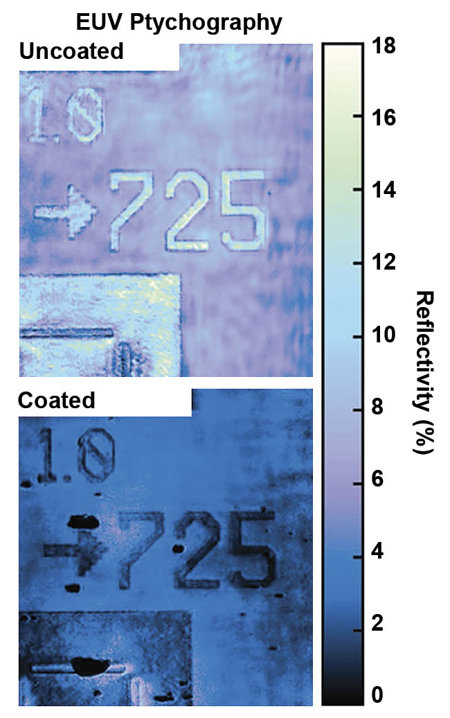
EUV ptychography performed on a patterned sample that is uncoated (top), and coated with 100 nm of aluminum (bottom). The partial transmissivity of the EUV beam through aluminum allows detailed characterization of interfaces and diffusion layers buried beneath the surface. Adapted with permission from E.R. Shanblatt et al., Nano Letters. See Reference 7.
The ultimate spatial resolution of the imaging method is not the size of the illumination spot, but rather it depends on the numerical aperture (NA) of the captured scatter, the illumination spot size, and the image sensor resolution. In ideal conditions, the resolution is D = 0.61 λ/NA,
and the illumination spot size must be <<D * Npixel, where Npixel would (for example) be 1024 for a 1024 × 1024 image sensor. An important aspect of ptychography is that neighboring spots have a large degree of spatial overlap, providing redundant information to the ptychographic reconstruction algorithm that is used to overcome the phase retrieval problem and recover complex wave profiles, including amplitude and phase information, for both the incident and transmitted/reflected beams.
Lensless imaging
Because of the lack of optics and associated aberrations, ptychography is known as a lensless imaging technique. In theory, it’s able to provide image resolution that is only a function of wavelength and numerical aperture, as dictated by diffraction-limiting conditions. Therefore, implementation of the technique with short wavelengths (on the order of 10 nm) is a hugely attractive avenue for high-resolution imaging.
This is especially true in the EUV to x-ray regime, where generally both the quality and the numerical aperture of imaging optics are severely limited. This has resulted in nearly an order of magnitude improvement in image resolution at any given wavelength, which has been transformational, particularly for the EUV. In the past, EUV image resolution was not particularly better than using visible light. Now, it is the highest-resolution tabletop-scale optical imaging technique.
Much of the early development and demonstration of CDI was done at synchrotrons1. But with recent advancements in tabletop short-wavelength coherent light sources, the CDI and ptychography methodologies have found increasing applications in laboratory settings2,3,4.
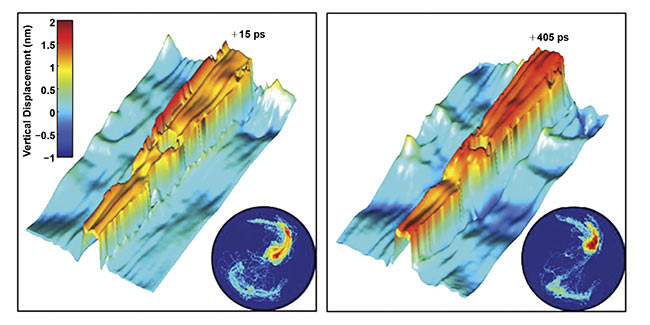
CDI imaging performed in a pump-probe arrangement to observe transient processes in a nickel nanoantenna on femto- to picosecond timescales. An IR pump was used to generate acoustic waves across the surface of the sample, which were observed with reflection CDI. Insets show 2D histograms of reconstructed
amplitude and phase. Adapted with permission from R.M. Karl et al., Science Advances. See Reference 8.
Various flavors of CDI have been developed — including transmission CDI, Bragg CDI, reflection CDI, and ptychotomography — to address different types of imaging problems and sample geometries5,6. Phase Focus Ltd., based in Sheffield, England, has pioneered the use of ptychography reconstruction in the visible wavelength range, using its wide-area scanning and unique ability to obtain phase information to provide contrast in biological science.
CDI ptychography in a reflection geometry — as pioneered by the JILA lab at the University of Colorado Boulder — has several attractive features. First, no (or minimal) sample preparation is necessary because measurements can be made on the surface of a large, bulk sample with no particular surface requirements such as conductivity or surface finish. This contrasts with soft x-ray imaging in transmission as implemented at synchrotrons or TEM/STEM (which needs thinned samples), or STM (which requires conductivity).
Amplitude and phase information in the reflected wave is highly sensitive to local properties of the material. Compositional variations are largely manifested in amplitude variations, as even small changes in molecular bonding or chemistry can cause attenuation differences at low energies — even more so if ptychography is intentionally performed with beam energy tuned to elemental absorption edges or known bonding energies. This provides excellent compositional sensitivity, particularly for low-Z materials, where complementary surface techniques such as SEM-based imaging and EDS/WDS (energy dispersive spectroscopy/wavelength dispersive spectroscopy) analysis have proved inadequate.
Additionally, the phase portion of the reconstructed image is closely correlated with topography of the surface, enabling relative height measurements at a small fraction of the wavelength of the light. And by performing imaging with different beam energy in the EUV to soft x-ray range, the microscopist can also tune the sensitivity of reflected light between surface and subsurface regions of the sample. This allows users to observe hidden features, defects, interfaces, or depth profiles that would not be detected with a purely surface-sensitive technique such as atomic force microscopy (AFM). Indeed, CDI with deep UV and extreme UV light combines aspects of SEM and AFM with new chemically sensitive contrast mechanisms and subsurface imaging capabilities7.
If implemented with ultrafast laser pulses as a light source, the reflection geometry is amenable to pump-probe schemes. In this arrangement, a pump laser pulse arrives just before the actual CDI measurement probe pulse and serves to inject energy into the material to perturb it, after which it begins to relax. By varying the time between pump and probe, the CDI measurements can then be stroboscopically made at different snapshots in time, yielding insight into dynamic events on femtosecond to picosecond timescales8.
The CDI reflection ptychography approach has begun to see applications in a variety of fields. In the physical sciences, CDI sits adjacent to complementary approaches such as SEM, AFM, and transmission x-ray microscopy, offering excellent nondestructive and noninvasive compositional and topographical imaging for materials science, chemistry, nanoscience, condensed physics, and semiconductor applications.
For biological specimens, CDI in the EUV spectral range has been used for high-resolution imaging of fixed, embedded, and unstained neurons using transmission CDI3. The relative ease of sample preparation — including the lack of heavy metal staining that would otherwise be required for high-resolution imaging using electron microscopy, and apparent lack of acquisition-induced sample damage compared with hard x-ray synchrotron microscopy — holds great promise for imaging life science samples as part of a correlative workflow.
Just as microscopy techniques such as confocal, scanning electron, and x-ray tomography began as experiments themselves and then gradually matured into routine techniques for the nonexpert user, the industry is on the dawn of a new range of possibilities based on coherent, ultrafast laser light sources. As the sources and associated detectors and software continue to advance and evolve into off-the-shelf complete solutions, new experiments based around nanometer spatial resolution and femtosecond time resolution will emerge to offer insights into previously unresolved properties and functions of materials and devices.
Meet the author
Will Harris is global business development manager at KMLabs Inc., where he helps grow the market of techniques based on ultrafast laser sources to a broader scientific audience. He has a Bachelor of Science degree and a doctorate in mechanical engineering from the University of Connecticut, with a background in engineering and materials science, focusing on high-resolution microscopy and spectroscopy techniques; email: [email protected].
References
1. B. Abbey et al. (2013). From grain boundaries to single defects: a review of coherent methods for materials imaging in the x-ray sciences. JOM, Vol. 65, Issue 9, pp. 1183-1201.
2. B. Zhang et al. (2015). High contrast 3D imaging of surfaces near the wavelength limit using tabletop EUV ptychography. Ultramicroscopy, Vol. 158, pp. 98-104.
3. P. Baksh et al. (2017). Quantitative evaluation of hard x-ray damage to biological samples using EUV ptychography. IOPscience, JPCS, Vol. 849, Conference 1, p. 012034. https://doi.org/10.1088/1742-6596/849/1/012034.
4. J. Rothhardt et al. (2018). Table-top nano-
scale coherent imaging with XUV light.
J Optics, Vol. 20, Issue 11, p. 113001.
5. J. Miao et al. (2015). Beyond crystallography: diffractive imaging using coherent x-ray light sources. Science, Vol. 348,
Issue 6234, pp. 530-535.
6. F. Pfeiffer (2018). X-ray ptychography. Nature Pho, Vol. 12, pp. 9-17.
7. E.R. Shanblatt et al. (2016). Quantitative chemically specific coherent diffractive
imaging of reactions at buried interfaces with few nanometer precision. Nano Lett, Vol. 16, Issue 9, pp. 5444-5450.
8. R.M. Karl et al. (2018). Full-field imaging of thermal and acoustic dynamics in an individual nanostructure using tabletop high harmonic beams. Sci Adv, Vol. 4, Issue 10, eaau4295.