George McNamara would like confocal microscopes to count.
When McNamara, image core manager at the University of Miami’s
Miller School of Medicine, says this, he isn’t talking about the importance
of the four confocal instruments he manages. Instead, he is talking about tallying
photons. That can lead to an accurate assessment of the number of fluorescing molecules
at a given spot in a tissue sample, thereby ushering in a fundamental research change,
McNamara believes. “This will turn cell biologists into single-molecule accountants.”
This ability to quantify what is going on in cells could have
a profound impact on research and the imaging core’s users. Today only 10
percent image live cells, but McNamara would like to see 90 percent doing live-cell
molecular-count imaging in 10 years.
Advances in technology, particularly the ability to resolve features
a few tens of nanometers on a side, may provide a means to achieve this. Along with
this trend toward greater resolution, other confocal microscopy developments involve
the incorporation of quantitative and interactive techniques. A third trend involves
faster and longer confocal image acquisition. But these developments are not without
cost, such as the need for new detectors and greater instrument stability.
The benefits of imaging smaller volumes rest on the fact that,
as dimensions shrink, so too do the number of molecules within a given space. When
the size approaches a box measuring perhaps 20 or 50 nm on a side, most locations
within a cell will contain a small and discrete number of fluorophores, which makes
counting easier.
“It’s either 0 or 1,” McNamara said of the simplest
case.
He acknowledges that distinguishing between 10 and 11 fluorophores
in these situations may be difficult. But, he predicts, being able to bin tiny volumes
into individual categories of up to perhaps 10 molecules would be enough to transform
research.
Achieving this type of performance could be done with recently
available fluorescence nanoscopes. As their name implies, such devices image on
the nanometer scale, capturing details from spaces much smaller than the micron-size
cubes imaged with conventional confocal microscopes.
One nanoscope implementation that is compatible with confocal
devices comes courtesy of Leica Microsystems Inc. of Bannockburn, Ill. The company
has licensed stimulated emission depletion (STED) microscopy technology from concept
originator and developer Stefan W. Hell, director of Max Planck Institute for Biophysical
Chemistry in Göttingen, Germany.
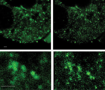
Comparison of confocal micrograph (left) with 240-nm X-Y resolution and stimulated emission
depletion (STED) microscopy nanograph (right) with 80-nm X-Y resolution using Alexa
Fluor 488. Top panels: 28-nm pixel size; bottom panels: cropped region zoomed 5x
using Adobe Photoshop CS5. Scale bar = 1μm. Images acquired by Charles Hemphill
of Leica Microsystems and George McNamara of the University of Miami. Specimen courtesy
of professor X. Mike Xu and Robert Moore, University of Miami.
The technique works by selectively switching off fluorophores,
with the achievable resolution determined by, among other things, the intensity
of the de-excitation beam. Hell noted that resolution of 5.8 nm has been achieved
– far better than the classical diffraction limit of hundreds of nanometers
for visible wavelengths.
This method is a natural fit with confocal microscopes because
it is a scanning technique. It also works with fluorescent proteins that are important
for live-cell imaging, enables the spatial arrangement of two molecular species
tagged with different fluorophores to be determined, and allows video-rate imaging,
Hell said.
“If integrated in a confocal microscope, STED allows one
to take 3-D images from the interior of living cells noninvasively. One is not limited
to imaging sample surfaces,” he added.
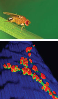
Top: The common fruit fly, Drosophila melanogaster. Bottom:
Adding another dimension can be done with confocal microscopy, as in this 3-D rendering
of a third-instar D. melanogaster larval neuromuscular junction. Bottom
image taken by Cheryl Herrera and processed by Jennifer Meerloo, University of California,
San Diego.
Chris Vega, Leica Microsystems’ product marketing manager
for confocal microscopy, noted that this superresolution is achieved optically during
the scan, without the need for postprocessing. This makes it possible for the company’s
products to image at speeds of up to 30 fps, to do so with multiple channels and
to achieve resolutions down to 50 nm.
Such an improvement is not free. For one thing, as imaging volumes
shrink, the number of sources and photons also drops. Going from a micron cube to
one that measures 50 nm cuts the number of photons by nearly four orders of magnitude,
everything else being equal. Thus, there either must be a more intense source, much
longer collection times, brighter fluorophores or better detectors.
The first option, Vega said, can damage cells and photobleach
fluorophores. The second slows throughput and requires extremely stable experiments.
The third is the subject of active research but is something Leica doesn’t
do. Consequently, the company is taking the fourth route and introducing a new type
of detector.
Vega noted that, traditionally, one detector option has been photomultiplier
tubes, which offer a wide dynamic range but lack sensitivity. The alternative has
been avalanche photodiodes (APDs), which present the opposite in benefits and drawbacks.
Leica now will offer a third choice.
“The hybrid detector, or HyD, basically provides the sensitivity
similar to an APD but with the dynamic range of a PMT [photomultiplier tube]. So,
basically, you get the best of both worlds,” Vega said.
The new detector has been demonstrated and will be available soon.
Other aspects of Leica technology also ensure that the greatest possible number
of photons is captured for imaging. In particular, Vega points to the use of acousto-optical
beamsplitters. Because this approach is filter-free, it minimizes photon losses
along the optical path.
Jennifer Meerloo, managing director of the shared microscopy center
at the University of California, San Diego, School of Medicine in La Jolla has implemented
superresolution using structured light. This approach extracts information from
distortions in a projected pattern of light and yields 3-D data. She believes that
there could be a place at the center for confocal-compatible superresolution and
would like to see other improvements.
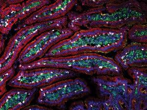
Mouse small intestine
labeled with fluorophores Alexa Fluor 546 (yellow), Alexa Fluor 488 (cyan-green)
and Alexa Fluor 647 (red) and imaged via confocal microscopy. Image taken by Jongdae
Lee, University of California, San Diego.
“If I had the capability on hand to increase the speed of
scanning without sacrificing optical sectioning, area or quality, we could do more
high-throughput and dynamic confocal work,” Meerloo said.
Brendan Brinkman, product manager of user scanning confocal microscopes
at Olympus America in Center Valley, Pa., noted that his company has tried to satisfy
this plea for speed in a number of ways. For example, some confocal microscopy applications
involve photoswitching and photoactivation of fluorophores. This is particularly
true for optogenetics, which combines optical and genetic techniques to trace neural
circuits.
Traditionally, in doing this, scanning systems would stop at a
point, activate the fluorophore and then collect the image. Data could be lost because
photoactivation could fade fairly quickly and switching was not instantaneous.
Olympus developed and now offers a simultaneous stimulation module
to attack this problem. In doing this, the company added another set of galvo mirrors
to the standard set found in all scanning confocal microscopes. This was combined
with other technology that enables any laser line to be split off and used for either
imaging or stimulation. Thus, a 488-nm line can be used for scanning, while at the
same time, a 405-nm line is used for stimulation.
“That allows us to, with one scanner, simultaneously image
and then, with this SIM scanner, photoactivate. We can do FRAP [fluorescence recovery
after photobleaching] experiments, we can do photoswitching experiments, all in
real time, so you don’t have any loss in data,” Brinkman said.
In the same vein of speeding things up and expanding capabilities,
the company also offers a host of other imaging and analytical methods that can
be added to its systems. These include multiphoton and coherent anti-Stokes Raman
scattering imaging as well as raster image correlation spectroscopy. The first two
methods allow intrinsic imaging of some biomolecules, eliminating the need for fluorophores.
Olympus also is working on the robustness of its optical components,
which leads to other benefits; e.g., Brinkman points to the company’s silicone
oil immersion objectives. Unlike other immersion objectives, their optical performance
doesn’t degrade over time. They also provide good refractive index matching
with tissue, minimizing optical loss and spherical aberration.
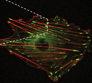
Confocal image of a vascular smooth muscle cell transfected with pGFP-vinculin and pmRFP-actin.
White dashed line represents functionalized atomic force microscope tip on top of
the cell, used to mechanically stimulate it and study cellular reactions. Courtesy
of Andreea Trache, Texas A&M University Health Science Center.
Because of that, these objectives allow the system to have a higher
numerical aperture, which helps lower the laser power used in scanning. That and
the refractive index matching enable live-cell experiments to run for a longer time.
These objectives, along with imaging and analytical capabilities
that provide quantitative live-cell data, can transform confocal systems, allowing
them to interrogate a cell for measurements as it is being imaged.
“We call it interactive imaging, where you’re getting
to actually interact with the live sample,” Brinkman said.
An example of an interactive cellular examination via confocal
microscopy can be seen in research from Andreea Trache of the Texas A&M Health
Science Center in College Station. Trache, an assistant professor of systems biology
and translational medicine, used an atomic force microscope tip coated with fibronectin
and a spinning disk confocal microscope to mechanically stimulate and image GFP-labeled
cells in real time, something not possible before.
She is studying how cells sense and adapt to mechanical forces
in their micro-environment, information that could prove useful in understanding
such diseases as hypertension and atherosclerosis. Trache reported on this protocol
in the October 2010 Journal of Visualized Experiments, with initial results indicating
that cells remodel themselves in reaction to certain stimuli.
Such research has required a high-sensitivity camera (from Tucson,
Ariz.-based Photometrics), a noise-free setup and plenty of patience, she said.
“We work at very low laser light intensity so we don’t photobleach the
cell. We have to keep the cell healthy during the period of the experiment, which
is pretty long. It’s 80 minutes.”