
DGC Helps Illuminate Mechanisms of Alcohol Dependency
Dodt gradient contrast microscopy offers a powerful means of imaging colorless specimens and can be integrated with patch-clamp studies.
By John Wingerd
Imaging transparent, colorless specimens has always presented a challenge in optical microscopy. In many applications, it is not desirable or even possible to use fluorescent dyes, stains or transgenic probes to render target objects more visible.
While differential image contrast (DIC), often in the form of the Nomarski microscope, is widely employed, its optical complexity can cause limitations when the contrast image is combined with some other measurement modality or imaging technique (such as patch-clamp measurements or optogenetic stimulation, or even simply epi-fluorescence).
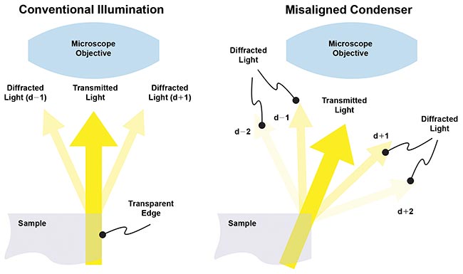
Figure 1. With conventional illumination, positive and negative diffracted sidebands are detected almost equally and their 180º phase difference cancels their amplitude. With oblique imaging, the positive (or negative) sidebands are preferentially collected by the objective. Courtesy of Siskiyou Corp.
The Dodt gradient contrast (DGC) method is an alternative proven to avoid these drawbacks and is being used by researchers to help explore the cellular basis for acquired alcohol dependency in the mammalian brain. Specifically, it enables critical positioning of patch-clamp electrodes in model (mouse) animal brains.
Electrophysiology, on the whole, enables the measurement and analysis of an extraordinary variety of dynamic electrical characteristics of excitable tissues. These can range from a large population of cells to an individual ion channel in a single patch of biological membrane from a single cell.
This latter type of measurement constitutes one of the most popular forms of electrophysiology and is typically referred to as whole-cell voltage clamp. With this, a glass micropipette acts as a microelectrode that is brought into gentle contact with the cell plasma membrane. Then a low level of suction draws the membrane into the aperture of the pipette and creates an ion-impervious seal between the rim of the pipette and the plasma membrane of the cell.
The discovery of the astonishingly strong “electrical seal” between the micropipette glass and biological membrane enabled Burt Sakmann and Erwin Neher at Munich’s Max Planck Institute to attain the requisite electrical recording fidelity to prove the existence of individual ion channels. The work earned them the 1991 Nobel Prize in Physiology and Medicine.
Typically, a brief pulse of suction is applied to the electrical seal to rupture the plasma membrane that separates the internal milieu of the electrode from the cell of interest, establishing an optimal low-resistance electrical circuit into the cell. This, in turn, enables electrical measurements to be made relative to a reference electrode located in the external buffer in which the sample is maintained.
In order to enhance the signal-to-noise ratio of signals as small as nano- or even picoamps, specialized circuitry clamps the voltage at a set value (typically the resting potential of -70 mV) and records the current required to maintain this voltage during transmission spikes and other events. The technique is usually referred to as patch-clamp measurement.
A critical task in patch-clamp experiments is positioning the electrode(s) — up to several electrodes are sometimes involved in complex stimulation and sensing experiments. Each electrode is usually positioned visually using multi-axis micromanipulators; DC servo systems are often preferred because they provide the highest stability and lowest electrical noise, in part because they draw no current when stationary. (For a more detailed discussion of patch-clamp techniques, see “Comparing Micromanipulators for Electrophysiology,” BioPhotonics, April 2015). The visualization required for this operation is challenging because the cells are usually colorless, as are the transparent glass electrodes.
Dodt gradient contrast
Most methods for imaging transparent objects rely on phase difference caused by refraction; the phase of transmitted light is retarded according to the refractive index of the object in its path. Various techniques use some type of interferometric method to convert phase differences into intensity differences so they can be sensed visually or digitally.
In the 1990s, professor Hans-Ulrich Dodt at the Max Planck Institute perfected an optically simpler method based on oblique imaging that relies on diffraction rather than refraction. When light strikes an edge with a refractive index discontinuity between two transparent materials (e.g., glass and saline), much of the light is transmitted with no deviation (Figure 1). However, some light is diffracted, with the particulars depending upon wavelength and the magnitude of the refractive index step.
The light diffracted in the positive orders is phase shifted by λ/4 (90º), and the light diffracted in the negative orders is shifted by -λ/4. In a conventional microscope used in transmission with on-axis illumination and viewing, both the positive and negative diffracted orders are detected almost equally and so cancel out in the image plane; the detector only “sees” the on-axis transmitted light.
In oblique imaging, the sample is illuminated with the condenser illumination misaligned in an off-axis direction. Now, only certain positive (or negative) diffracted orders are preferentially detected. Because this light is shifted λ/4 from the undiffracted light, interference occurs, causing the edge to appear as dark (or light) in the image plane.
In simple oblique imaging, only edges that are perpendicular to direction of illumination shift show contrast (i.e., a θx misalignment of the illumination will highlight edges with a component in the y direction). The misaligned condenser has to be rotated to sequentially “see” all the edges in the sample. Dodt modified the illumination method so that a wider angular range of edges can be seen with reasonable contrast. In practice, however, operators still often rotate the condenser to provide a slight increase in visibility for targeting image features.
DGC, also called amplitude image contrast (AIC), uses a diffuser and a condenser with a curved (annulus) mask that spans one quarter of the condenser circumference (Figure 2). This results in an off-centered illumination profile with a smooth intensity gradient in two perpendicular directions. Any transparent object with edges that diffract light will always be visible, irrespective of the condenser angular orientation. The diffuser also ensures that there is non-zero light intensity across the entire condenser field, making the Dodt images very similar to normal images of the sample, albeit with highlighted dark and light edges.
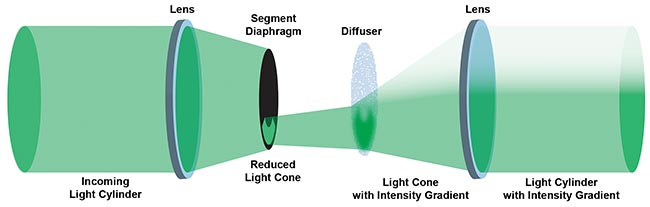
Figure 2. This schematic illustration of the Dodt-type condenser shows how the illumination beam is created with an intensity gradient. Courtesy of Siskiyou Corp.
Several suppliers of microscope components and accessories provide a fully integrated DGC condenser that is compatible with most commercial upright and inverted microscopes. An example is the Siskiyou IS-GCI, which is available with or without an integrated LED source(s).
In comparison, DIC microscopy, often called Nomarski interference contrast (NIC) or Nomarski microscopy, is the mostly widely employed refraction-based approach for imaging transparent objects. It uses Wollaston prisms after the condenser to create linearly polarized wavefronts of equal intensity that are orthogonally polarized to each other. The arrangement introduces a slight offset or shear in the two wavefronts in the sample plane.
After passing through the sample, the two wavefronts are recombined in the infinity space of the microscope using another pair of Wollaston prisms. Every point in the image is thus a result of interference between light from a point in the sample and light from a point that is slightly offset. This produces light and dark areas in the final image, where steeper edges produce higher contrast.
While DIC often produces superior images to DGC, the latter is widely used, particularly in patch-clamp studies, for several reasons. The first is cost: Wollaston prisms use expensive calcite, often requiring crystals with a large, clear aperture free of striations, cracks and other defects. The second advantage is the simplicity of DGC, which provides multimodal compatibility.
At the University of Texas, the electrophysiology lab of Richard Morrisett, a professor in the College of Pharmacy, is equipped with multiple optical microscopes, including several based on the Siskiyou MRK100 modular platform. “We often can get higher-quality images with a Nomarski configuration, but a primary application for us is patch-clamp setup,” he said. “Here we need to view ‘spiny’ neurons measuring <10 µm in diameter, as well as one or more glass micropipettes with tip diameters around 1 µm. The DGC method produces images that are usually higher quality than we need for this purpose.”
Morrisett added that the lab increasingly combines patch-clamp measurements with other modalities, particularly epi-fluorescence imaging, and also runs experiments with more than one patch-clamp. They are also starting to use optogenetic tools for neural stimulation, which requires introducing a focused light beam into the sample. DIC would pose a big problem, because DIC imaging requires dedicated optical components. Their solution, said Morrisett: “We insert a commercial optogenetic illumination module and LED illumination into the microscope’s infinity space for photoactivation of neurons. DIC requires optics in the infinity space, which would have to be somehow removed without disturbing the alignment of the microscope and this optogenetic stimulation light.” He adds that DIC also would make things unacceptably congested for experiments using multiple electrodes.
For several years, the focus of Morrisett’s research has been to unravel the neurological basis of alcohol dependency and craving based on the mouse model. Specifically, his group is studying the mechanisms by which ethanol modulates information processing in mesocorticolimbic structures in the murine brain. Researchers induce alcohol craving and dependency in groups of lab animals by exposing the mice to alcohol vapor for several days each week, following the chronic intermittent ethanol (CIE) exposure method, developed by professor Howard Becker at the Medical University of South Carolina.
The animals have two drinking sources available to them: distilled water and a solution that contains 15 percent ethanol. After a few weeks, the animals develop a significantly increased preference for the ethanol solution; they drink 50 to 100 percent more alcohol than nontreated animals (Figure 3). If the mice are subsequently deprived of an alcohol source, and no longer subject to breathing ethanol vapor, after a few more weeks, they lose interest in any offered alcohol. The group also uses a strain of mice that are genetically predisposed to drink alcohol.
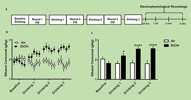
Figure 3. This timeline shows that chronic intermittent ethanol (CIE) exposure induces increased voluntary ethanol consumption in the lab mice. It also shows the timeline of the electrophysiological recordings summarized in figure 4. Courtesy of Renteria et al./Addiction Biology.
At several stages, starting with time zero as a baseline control, some of the mice are humanely anesthetized and killed, and brain slices that are approximately 220 to 250 µm in thickness are placed in a microscope slide in continuously oxygenated artificial cerebrospinal fluid (ACSF). They are then probed using patch-clamp technology. (As noted earlier, the group also sometimes incorporates fluorescence microscopy, and has recently added optogenetic stimulation to the experimental mix, using mice expressing tdTomato.)
“The most exciting part of our research is that we can see plasticity changes — changes in the neurons’ ability to signal each other — that are induced and dissipate on this same timescale as their consumption pattern,” Morrisett said. “We can finally see the cellular basis for alcohol craving in the mammalian brain.”
Morrisett’s group is focused on spiny neurons in the nuclear accumbens (NAc), which is a major target of the ventral tegmental area (VTA). “Multiple brain locations are involved with alcohol craving and consumption,” he said. “This includes initial information from the olfactory/taste processing region, feedback from the VTA where ‘feel good’ sensations are handled, memories from other limbic structures encoding memories of prior rewards, or stressors and anxiety associated with rewards, as well as conscious brain areas [that] control executive functions emanating from the prefrontal cortex — ‘Should I drink more of this or not?’”
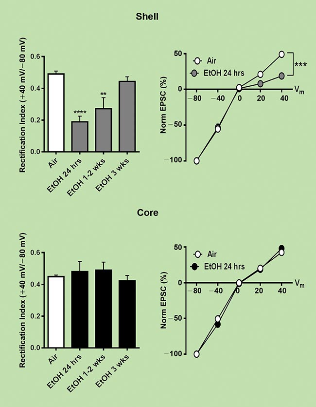
Figure 4. Normalized patch-clamp electrical data for the D1 neurons in the NAc shell and D2 in the core. The synaptic activity of the D1 neurons shows changes that correlate with the alcohol exposure, whereas the D2 neurons show no such changes. Courtesy of Renteria et al./Addiction Biology.
Their experiments have shown that there are actually two types of dopamine receptors in the NAc — so-called D1
and D2 receptors — where D1 receptor-expressing neurons located in the NAc shell are associated with positive feedback, and D2 receptor-expressing neurons in the NAc core are associated with negative aspects of reward processing and feedback.
Moreover, they have observed glutamatergic-mediated transmission onto the D1 type changes relative to that of the D2 group, contemporaneous with the induced craving (glutamate is the principal excitatory neurotransmitter in the brain). As the craving dissipates, they have seen this return toward normal levels.
“Sadly, alcoholic addiction is a worldwide problem with huge individual and societal consequences,” Morrisett said. “Understanding the cellular and chemical basis for alcohol craving can hopefully eventually lead to improved treatments for this often debilitating condition.”
Patch-clamp studies are yielding detailed information about intracellular processes, enabling scientists to unravel many of the specifics of how various biochemical processes operate. But a prerequisite for this work is an imaging modality that is compatible with patch-clamp instrumentation, including its required high-precision micromanipulators. DGC has proven to be just such a method.
Meet the author
John Wingerd is a senior mechanical engineer at Siskiyou Corp. in Grants Pass, Ore.; email: johnw@siskiyou.com.
/Buyers-Guide/Siskiyou-Corporation/c16507