The use of diffuse optical spectroscopy and accompanying methods has been shown to capture, in real time, diseases such as breast cancer that may be harder to identify with other technologies.
Diffuse optics and the study of light absorption and scattering in various tissues can track blood oxygenation and other factors that help assemble an accurate medical diagnosis. Light scatters multiple times when traveling through a thick tissue sample, hence the “diffuse” property of this technique. Diffuse optical imaging has been shown to be a reliable measure for evaluating the health and outcomes of patients undergoing cardiac surgery, metabolic treatment, and tests for cancer (Figure 1). Breast cancer, in particular, has been both a tangible and virtual target for these techniques.
Figure 1. Cancer cells among red blood cells. Courtesy of iStock.com/CreVis2.
Diffuse optical spectroscopy (DOS) characterizes tissue with the aid of low levels of light in the red to near-infrared range. Light is emitted in pulses or continuous waves, and photons are collected by a detector after they have passed through the tissue. By contrast, diffuse correlation spectroscopy (DCS) measures changes in scattering of light as they relate to moving objects, such as red blood cells, and provides information about blood flow.
A variety of research institutions and companies specializing in optical technologies have set to work creating DOS and DCS systems in recent years. Many of these systems have allowed for the creation of hand-held, low-impact equipment that can be brought to the bedside by a physician.
An image of breast cancer
One of the most significant studies in this area was called the ACRIN 6691 trial. It was carried out between 2011 and 2013, and involved 60 female breast cancer patients at seven institutions. The results were published in 2016. The project focused on the value of diffuse optical imaging for neoadjuvant chemotherapy, otherwise known as preoperative systemic therapy, which aims to identify and contain cancer, making later operations, radiation, and drug treatments more effective. Measurements were taken with a probe, using frequency domain and broadband NIR (650 to 1000 nm) spectroscopy. This range helped tabulate light absorption and scattering properties of oxyhemoglobin, deoxyhemoglobin, water, and lipids, which are the primary components found in the human breast. The levels of these elements change significantly with the growth of tumors1.
DOS imaging scans were taken prior to the start of therapy, five to 10 days after the first cycle of treatment, midway through the regimen, and then within three weeks after the therapy but prior to surgery. Researchers found that while responses to treatment varied, this method showed that the tissue optical indexes (TOIs) obtained during the study strongly determined where and how a positive outcome would result1.
One researcher who took part in this study was Thomas O’Sullivan, an assistant professor of electrical engineering at the University of Notre Dame. He said DOS is most promising in a clinical setting when used in combination with traditional medical imaging, such as ultrasound and MRI. But he added that the use of DOS is sometimes preferred over other technologies due to its relatively low cost and readability of the results (Figure 2).
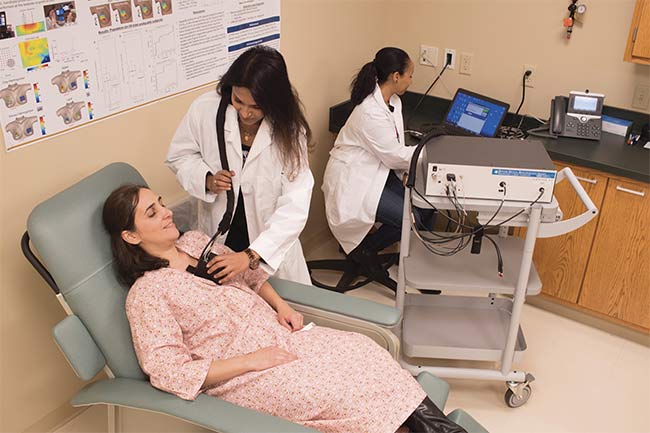
Figure 2. Simulated breast measurement using a diffuse optical spectroscopy (DOS) imaging system in the laboratory of Thomas O’Sullivan. Courtesy of Wes Evard/University of Notre Dame College of Engineering.
Last year, O’Sullivan and several other researchers published a study assessing the use of DOS in investigating the physiology of human lactation. They asserted that the technique could establish changes in mammary tissue composition during the process of involution, where the lactating breast returns to its prepregnant state. The participant in the study was a 37-year-old woman who O’Sullivan said had taken part in studies before and had already undergone chemotherapy in her right breast. Her other breast was examined every one to three weeks, for a total of nine times, with an apparatus (developed by the team) that contained a frequency domain photon migration module with fiber-coupled, modulated laser diodes at four wavelengths — 660, 690, 780, and 830 nm (the same technology used in the ACRIN 6691 trial)2.
During the course of the study, lipid content in the nonareolar region (outside the pigmented area surrounding the nipple) increased while water and hemoglobin decreased (Figure 3). Significant oxygen and water content changes were noted as well. The authors of the study concluded that DOS was a safe and effective way to measure changes in the human breast during the lactation process2.
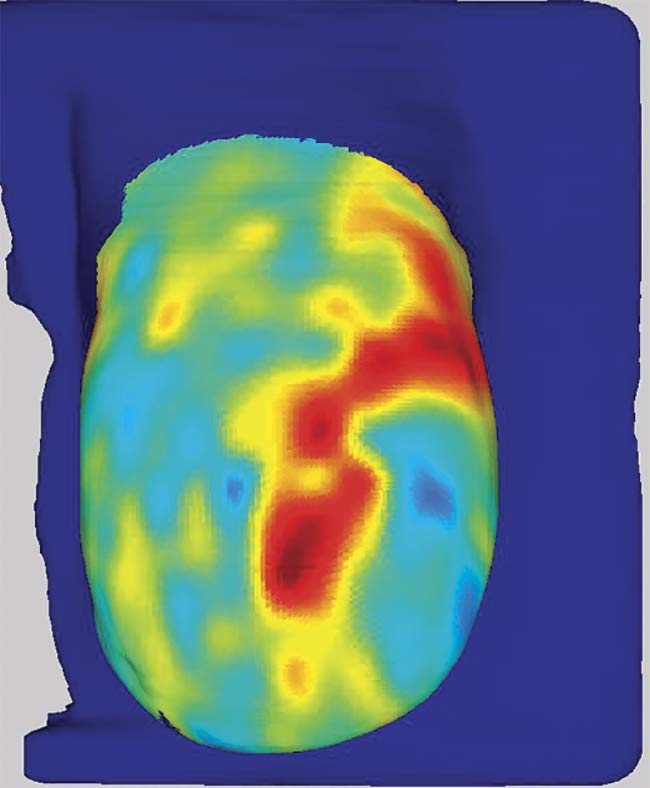
Figure 3. A DOS image of oxyhemoglobin concentration in a healthy breast. Courtesy of Thomas O’Sullivan.
O’Sullivan said the implications of these findings were significant because of limitations in the detection of health risks during pregnancy.
“MRI, for example, does not work in the lactating breast and cannot capture important imaging information, due to the water and blood dynamics,” he said. “We are currently working to make the technology more accessible by reducing size and cost, while also improving performance. A hand-held prototype is in development and has the potential to measure different kinds of treatment.”
Wearable monitor
A group of researchers led by Darren Roblyer, an associate professor in the Department of Biomedical Engineering at Boston University, has been developing a wearable diffuse optical probe to monitor the hemodynamics (variations in blood concentration) in breast cancer tumors (Figure 4). Changes in the vasculature of these growths has been directly linked to the progression of cancer. Samuel Spink, a doctoral student who works with Roblyer, discussed the hopes the group had for the probe at SPIE Photonics West in San Francisco earlier this year. Spink said the probe uses continuous-wave near-infrared spectroscopy (CW NIRS) to quantify these measurements. This work is valuable because in approximately 31% of cases where breast cancer patients are treated with neoadjuvant chemotherapy, the cancer remains3.
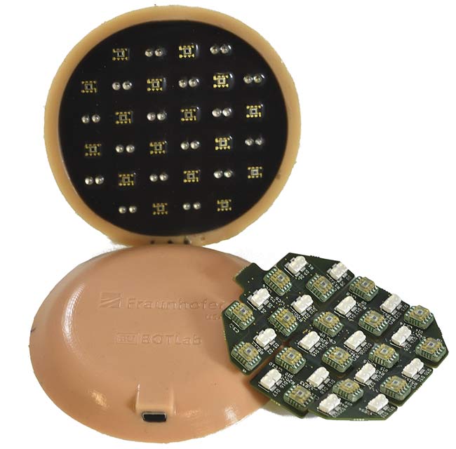
Figure 4. Two sides of a wearable DOS probe, and a printed circuit board that will be encased in silicone. Courtesy of Matthew Applegate.
“It is similar to a number of other wearable devices that utilize NIRS technology, which involves the illumination of tissue with at least two wavelengths in the near-infrared region, and subsequent detection of reflected light,” he said. “The simplest forms of this kind of probe employ a limited number of light-emitting elements, or sources, and light-detecting elements, or detectors. Our probe is unique in its number of source-detector pairs, or optodes. Our wearable probe has 32 sources and 16 detectors, producing 512 unique channels, and this number of channels provides enhanced spatial information, both laterally and in depth, when compared to wearable devices with fewer optodes.”
Chromophores that are targeted by these probes include oxyhemoglobin and deoxyhemoglobin. Spink said the light sources at the 750- and 850-nm range are commonly used for highlighting these chromophores. The probe was placed on a silicon optical phantom, into which a solution had been injected, to measure spatial resolution. In time, human patients will be monitored during paced breathing.
“We have yet to measure breast cancer patients with this wearable probe — only healthy volunteers — but we have approval to measure in the clinic and will be quantifying the hemodynamic response to paced breathing,” Spink said. “We believe that this response will vary in cancerous versus healthy tissue, so we hope to quantitatively differentiate between breast tumors and surrounding healthy tissue with paced breathing metrics.”
Like O’Sullivan, Spink said his research team believes that by fine-tuning this technology, they will be able to provide it to clinicians, who will be able to monitor patient response to neoadjuvant chemotherapy and personalize a treatment regimen.
He noted that while other wearable devices measure hemodynamics in the brain and muscles, his team focused on the breast.
The use of DOS, specifically for measuring the effectiveness of neoadjuvant chemotherapy in locally advanced breast cancer, was also the subject of a 2017 paper produced by a group of scientists that included Dr. Gregory Czarnota, a radiation oncologist at Sunnybrook Research Institute and a professor in radiation oncology and medical biophysics at the University of Toronto. The group positioned the patients and used a commercially available tomographic DOS imaging device produced by SoftScan in Montreal, examining 37 patients who were diagnosed with locally advanced breast cancer. Four diodes emitted near-infrared light at 690, 730, 780, and
830 nm. Optical collimators collected the photons that had passed through the patients’ breasts4.
The researchers concluded, after using a series of classification algorithms, that the technology effectively mapped the homogeneity of breast tissue — like Spink’s group, the measurement focused on such elements as oxyhemoglobin and deoxyhemoglobin — and also successfully evaluated the texture of tumors. Since neoadjuvant chemotherapy is intended to medically establish and limit the boundaries of cancer prior to a regular regimen of treatment, the researchers said an imaging system that can determine response would go a long way to track a successful outcome.
“DOS works well in a neoadjuvant setting,” Czarnota said. “We have known that there are early changes in tumor vasculature in the early stages of breast cancer, and with direct measurements of oxyhemoglobin and deoxyhemoglobin, we can produce a 3D image that shows the degree of heterogeneity and any changes that have occurred. Not all tumors respond the same to treatment, and with the use of DOS in conjunction with ultrasound and photoacoustics, we can also determine what is benign and what is malignant.”
Czarnota said, however, that one of the challenges to the commercial success of DOS systems is that companies producing the technology try to tie themselves to near-universal modalities such as mammography, which is part of mainstream medical treatment.
“Mammography is so established that when manufacturers try to tie themselves to it, there isn’t much commercial opportunity there,” he said. “But neoadjuvant chemotherapy is a procedure where DOS can find an opening and have a real impact.”
Also working on a DOS probe is a team from Simon Fraser University (SFU) that includes Majid Shokoufi and Farid Golnaraghi of the School of Mechatronic Systems Engineering. Citing a desire to reveal cancer characteristics as soon in the disease’s development as possible, the research team produced a diffuse optical breast scan (DOB-scan) probe that consisted of multiwavelength encapsulated light-emitting diodes (eLEDs) that operate in four NIR wavelengths: 690, 750, 800, and
850 nm. A linear array detector with 2048 sensors measured
the intensity of the back-scattered photons5.
The probe was initially used on phantoms (Figure 5) composed of a cylindrical acetal resin rod along with other substances to mimic the components in a human breast — water, lipids, oxyhemoglobin, and deoxyhemoglobin, with the last two producing greater contrast in cancerous lesions. While the probe has limited depth range at this point, due to separation between the light source and the detector, it has shown itself to be a promising tool to capture anomalies created by cancerous tumors during the course of neoadjuvant chemotherapy5.
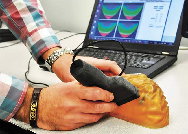
Figure 5. The SFU team’s device uses low-frequency light emitted from a hand-held probe to detect the presence of cancerous tissue in the breast (testing on a phantom). Courtesy of Simon Fraser University.
More recently, the probe has been used on human subjects and has received ethical approval for use on breast cancer patients, Shokoufi said. Both manual and automated software was used to tabulate data from the images, which met the specific needs of the researchers. The DOB-scan device itself was designed in-house by the research team.
“Four main chromophores are inside breast tissue, and we are looking for concentration images for these four chromophores,” Shokoufi said. “This is the reason why we have four different wavelengths. … Subjects will be asked to lie down in a supine [face up] position. The skin to which the probe will be attached will be cleaned with alcohol prep pads. The probe will be placed on the breast tissue where it needs to be scanned.”
He said that this examination has continued and the team is in the second phase of its clinical study, with an eye toward eventual application in a variety of settings. Shokoufi believes that as the technology and equipment are developed, more uses will be found.
“It can be used for other types of cancers, like prostate, but the probe shape and sensors must be redesigned,” he said.
References
1. B. Tromberg et al. (Oct. 15, 2016). Predicting responses to neoadjuvant chemotherapy in breast cancer: ACRIN 6691 trial of diffuse optical spectroscopic imaging. Cancer Res, Vol. 76, Issue 20,
pp. 5933-5944.
2. N. Bosschaart et al. (May 2019). Diffuse optical spectroscopic imaging for the investigation of human lactation physiology: a case study on mammary involution. J Biomed Optics, Vol. 24, Issue 5, p. 5006.
3. Early Breast Cancer Trialists’ Collaborative Group (EBCTCG) (2018). Long-term outcomes for neoadjuvant versus adjuvant chemotherapy in early breast cancer: meta-analysis of individual patient data from ten randomized trials. Lancet Oncol, Vol. 19, Issue 1, pp. 27-39.
4. W. Tran et al. (2017). Predicting breast cancer response to neoadjuvant chemotherapy using pretreatment diffuse optical spectroscopic texture analysis. Br J Cancer, Vol. 116, Issue 10, pp. 1329-1339.
5. M. Shokoufi and F. Golnaraghi (2019). Handheld diffuse optical breast scanner probe for cross-sectional imaging of breast tissue.
J Innovative Opt Health Sci, Vol. 12, No. 2, 1950008.
Focusing on Components for Measurement
A variety of companies produce technologies that provide valuable components for diffuse optical spectroscopy (DOS) and diffuse correlation spectroscopy (DCS) systems.
For example, ISS Inc. makes a fast 8-channel correlator that is used in applications of DCS, as well as in a module to capture measurements of the correlator itself and up to eight detectors. The system is used to monitor the consumption of oxygen in a patient’s tissue, along with blood flow. The corresponding software has also been produced by ISS.
“Our MetaOx is a combined FD NIRS [frequency domain near-infrared spectroscopy] and DCS instrument, all into one functional and portable unit,” said Beniamino Barbieri, president of the company. “The first units of the MetaOx have been purchased by various hospitals specializing in treatment of children.”
He said the main application for which the system is used is to monitor cerebral oxygen metabolism in children suffering from heart failure, although some researchers have begun using it to study sport physiology as well.
A European Connection
While a variety of research institutions in the U.S. and Canada seek a marketable solution for a swift and relatively inexpensive imaging technique to diagnose breast cancer, the SOLUS project has been underway for the last few years in Europe. The stated mission of this collaboration that unites corporate, collegiate, and governmental resources from several nations is to create a small photonic module (or a hand-held optode probe). The device will utilize both diffuse optical tomography — which uses diffuse optics to produce 3D images of tissue — and ultrasound/shear-wave elastography, which uses sonic waves to determine the elasticity of tissue, often a telltale sign of the presence of cancerous growths. Similar to counterparts developed in North America, the device is intended to measure water, collagen, and lipids, as well as blood characteristics.
“We combine two ultrasound techniques (B-mode scanning, which measures morphology, and shear-wave elastography) available commercially from SSI, partner of the project, with diffuse optical tomography,” said Paola Taroni, SOLUS coordinator and professor at Politecnico di Milano in Italy. “For the latter, all major components (picosecond pulsed laser driver, time-gated detector with a wide active area, and acquisition electronics) were developed within the project, as we need to combine high performances with very small size, and nothing was available commercially.”
According to Taroni, data is collected in a sequential manner, although B-mode ultrasound — which provides 2D display images composed of dots representative of echoes — works with optical data collection to provide spatial information. Currently, compiling test results takes several minutes. But assuming there is validation at the end of the project, a more efficient analysis, perhaps cloud-based, could be produced.
Researchers who are invested in the project point out that SOLUS is expected to significantly improve outcomes in health care settings that are currently limited to other modalities. While mammography is an effective tool in establishing the existence of growths, it often takes time-consuming and invasive procedures, such as biopsies, to determine whether cancers are malignant. In the meantime, of course, a sizable cost to both the individual and the health care system as a whole is incurred.
“The idea is to use the multimodal SOLUS device after positive screening mammograms, aiming at reducing noninvasively the number of needless false positive biopsies that are presently performed,” Taroni said. “So, I would say hospitals and diagnostic centers offering screening mammography programs will use this device.”
SOLUS is a broad-based initiative of the Photonics Public-Private Partnership of Photonics 21, and has been funded by the European Union’s Horizon 2020 research program. Taroni said many of the groups and institutions that are taking part in the project have collaborated with her research in the past, while others offered particular expertise in literature, so she chose them.
A statement from the project website reads, “There is a clear unmet need for a noninvasive diagnostic tool to achieve a more specific breast diagnosis and improve the quality of life of more than a million European women every year. … The new SOLUS system will allow more effective treatment and management, novel and improved therapy response prediction and monitoring, [and] enable personalized decision-making and therapy planning, and optimization for each patient.”