Christoph K. Hitzenberger, Medical University of Vienna
The development and application of optical
techniques and tools for biomedical imaging, diagnostics and therapy have been continuously
expanding for several decades. These advances are based on multidisciplinary efforts
requiring contributions from various fields such as physics, chemistry, biology,
engineering, informatics and medicine. While physicists explore the application
of basic optical principles to biomedical problems, and chemists develop new molecular
and target-specific probes, hard- and software engineers translate these results
into practical devices that are tested by clinicians for diagnostic and therapeutic
usefulness to patients. Based on the results, the clinicians can provide valuable
feedback to basic scientists and engineers for future improvements of their methods.
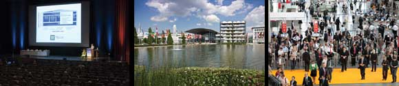
The European Conferences on Biomedical
Optics will be a highlight of this year’s Laser World of Photonics trade fair.
The show, held every other year, brings together key industry and research players
from around the world to spark growth in both sectors. Images courtesy of Messe
München GmbH.
To be successful, this multidisciplinary approach requires cooperation,
communication and exchange of ideas between scientists of the various involved disciplines.
The European Conferences on Biomedical Optics (ECBO), the largest group of biomedical
optics conferences in Europe, is a venue for this kind of communication and idea
exchange. The meeting is held every other year and is jointly sponsored by SPIE
and OSA. It is colocated with the world’s number one fair on lasers, optics
and optoelectronics, Laser World of Photonics in Munich, Germany, as well as with
numerous other society meetings organized by WLT, EOS, OSA, CLEO-Europe, IEEE/LEOS
and EPS.
ECBO 2011 comprises seven conferences:
• Advanced Microscopy Techniques II
• Clinical and Biomedical Spectroscopy and Imaging II
• Diffuse Optical Imaging III
• Molecular Imaging III
• Novel Biophotonic Techniques and Applications
• Optical Coherence Tomography and Coherence Techniques V
• Medical Laser Applications and Laser-Tissue Interactions V
A total of 458 contributions were submitted to these seven conferences,
an increase of approximately 13 percent over the figure from the previous event
in 2009. But ECBO not only strives to grow in quantity but also seeks to maintain
and further improve the quality of presentations. Toward this end, a full peer review
process was held by the program committee of each conference. This procedure was
first implemented at ECBO 2009.
Special events
The following special events of ECBO should not be missed:
There will be two plenary lectures on Tuesday, May 24:
• On the occasion of the 20th anniversary of OCT, Wolfgang Drexler, Medical University
of Vienna, will present a talk on “Twenty Years of Optical Coherence Tomography: Where Is It Heading?
• Mary-Ann Mycek, University of Michigan, will speak on “Optical Spectroscopy for Clinical Detection of Pancreatic Cancer.”
E-CLEO
Two joint sessions with E-CLEO are scheduled:
• On Monday, May 23, Jim Fujimoto, MIT, will give a tutorial on OCT, and Stefan M. Witte, Vrije Universiteit Amsterdam, will speak on “Label-Free Live Brain Imaging with Third-Harmonic Generation Microscopy.”
• On Tuesday, May 24, Emmanuel Beaurepaire, Ecole Polytechnique, will speak on “Nonlinear Microscopy of Tissues and Embryo Morphogenesis” Fiorenzo Omenetto, Tufts University, will present a talk on “Silk – New Opportunities in Optics and Photonics for an Ancient Material,” and Brian E. Applegate, Texas A&M
University, will speak on “Development of Transient Absorption Ultrasonic Microscopy.”
The postdeadline/hot topics session on Wednesday, May 25, will
feature late-breaking scientific results.
Technical presentations
Apart from these special events, the main attractions at ECBO
are the more than 450 presentations (talks and posters) submitted by authors from
all over the world. An overview of all the scheduled presentations would be beyond
the scope of this article. Furthermore, the diversity of studies (theoretical, modeling,
basic experimental, clinically oriented) makes it difficult to select the presentations
to be mentioned. Therefore, I have decided to give a short overview of some topics
and talks that received the highest scores in the peer review process, that are
likely to have high impact, and that seem to be most attractive to a general audience
with an interest in biomedical optics. (I know that especially the latter criterion
is rather subjective, and I apologize for any omissions.)
Among the most highly profiled presentations of the Advanced Microscopy
Techniques Conference are 3-D fluorescence lifetime imaging techniques for imaging
small (mm) and large (cm) samples (J. McGinty et al, Imperial College London); 3-D
imaging by self-interference fluorescence endoscopy (M. de Groot et al, VU University
Amsterdam); quantitative imaging of cell division in zebra fish embryos by multiharmonic
microscopy (N. Olivier et al, Ecole Polytechnique, CNRS, INSERM, Palaiseau, France);
and imaging of embryos with multiphoton light sheet microscopy (W. Supatto et al,
California Institute of Technology, Pasadena).
Some of the highlights of the Clinical and Biomedical Spectroscopy
and Imaging Conference are Raman microspectroscopy applications for identification
of bacteria (P. Rösch et al, Friedrich Schiller Universität Jena) and
for studying live cells in collagen matrices (F. Bonnier et al, Focas Research Institute,
Dublin); a new optical fiber sensor for pH measurement in the stomach (F. Baldini
et al, IFAC-CNR, Sesto Fiorentino, Italy); differentiation of melanin types by transient
absorption microscopy, which is potentially useful for melanoma diagnostics (M.C.
Fischer et al, Duke University, Durham, N.C.); and assessment of breast tumor margins
by quantitative spectral imaging (B. Yu et al, Duke University).
Breast imaging is also one of the highlights of the Diffuse Optical
Imaging Conference (P. Taroni et al, Politecnico di Milano). This work investigates
the replacement of hazardous x-ray mammography by nonhazardous optical radiation.
Other highly scored contributions to this conference include brain imaging and measurements
by functional near-infrared spectroscopy (NIRS) to study motor tasks (A. Torricelli,
Politecnico di Milano), cognitive tasks (A. Jelzow et al, Physikalisch-Technische
Bundesanstalt, Berlin), and the physiologic origin of task-evoked systemic artifacts
in functional NIRS (E. Kirilina, Freie Universität Berlin).
Among the most interesting presentations of the Molecular Imaging
Conference are a talk on sensing rare fluorescently labeled circulating cells in
vivo (M.J. Niedre et al, Northeastern University, Boston); other highlights of this
conference are focused on optoacoustic imaging of cancer receptors targeted by gold
nanorod conjugates (A.A. Oraevsky, Tomo-Wave Labs and University of Houston), on
structure and biomarkers in mice by multispectral optoacoustic tomography (A. Buehler
et al, Technische Universität München); and on the use of plasmonic nanoparticles
for molecular-specific cancer imaging in vivo (K.V. Sokolov et al, University of
Texas at Austin).
The Novel Biophotonic Techniques and Applications Conference covers
various techniques from diverse fields of biomedical optics. Top-ranked contributions
include monitoring of cell death and ionic homeostasis with digital holographic
microscopy (N. Pavillon et al, EPFL, Lausanne); in vivo evaluation of coronary plaques
by intravascular laser speckle imaging (S.K. Nadkarni et al, Massachusetts General
Hospital, Boston); intravascular photoacoustic imaging of human coronary atherosclerosis
(K. Jansen et al, Erasmus MC, Rotterdam); and Raman active phospholipid gold nanoparticles
for lung cancer detection (N.C.M. Tam et al, University of Toronto).
Highly profiled talks in the Optical Coherence Tomography and
Coherence Techniques Conference comprise novel Doppler OCT techniques (i) employing
a two-beam technique for microvasculature imaging of the human retina (S. Zotter
et al, Medical University of Vienna), and (ii) using a 1-μm center wavelength
for imaging blood flow deep in the choroid (Y.J. Hong, University of Tsukuba, Japan);
ultrahigh-speed retinal OCT beyond 1-MHz A-scan rate (T. Klein et al, Ludwig Maximilians
Universität München); high-speed functional OCT with extended focal depth
by using a Bessel beam (C. Blatter et al, Medical University of Vienna); simultaneous
800- and 1060-nm retinal OCT with retinal tracking (B. Povaay et al, Medical University
of Vienna); and dark-field optical coherence microscopy for functional cell imaging
(C. Pache et al, EPFL Lausanne).
Finally, some of the highlights of the Conference on Medical Laser
Applications and Laser-Tissue Interactions are presentations on how gold nanorods
illuminated by pulsed lasers can be used to modify or destroy cells (F. Rudnitzki
et al, Universität zu Lübeck); and on novel infrared optical probes for
detecting sentinel lymph nodes (F. Tellier et al, Université de Strasbourg/CNRS).
In addition, several papers directly related to clinical laser applications, clinical
aspects and results of clinical studies will be presented that should be of special
interest to the medical community.
Students welcome
Although there is an impressive breadth of high-quality presentations
scheduled, and the number of submissions to ECBO is still growing, ECBO is aware
that it has to take measures to attract future generations of scientists to the
field of biomedical optics. Therefore, we especially encourage students to attend
ECBO. To underline the importance of participation by future generations of scientists,
awards for outstanding contributions by students will be presented: the Toptica
Best Student Paper Award and the Toptica Best Student Poster Award (€1000 each).
Furthermore, student grants for attending the meeting will be sponsored by Thorlabs,
OSA and SPIE. These awards and, of course, the excellent opportunities to see the
latest scientific results and discuss and network with colleagues, will help to
attract talented students to our field and secure a bright future for biomedical
optics.
Meet the author
Christoph K. Hitzenberger is a professor and faculty member at
the Center for Medical Physics and Biomedical Engineering at the Medical University
of Vienna, Austria, and is co-chair of the European Conferences on Biomedical Optics;
e-mail: [email protected].