MÜNCHEN, Germany, March 7, 2022 — Researchers at the Fraunhofer Institute for Toxicology and Experimental Medicine (ITEM) have developed a procedure for characterizing immune cells in the lungs. The characterization method uses a technique called chip cytometry, and it offers advantage over existing techniques by enabling patient samples to be stored under refrigeration for months and examined for further parameters should new issues become relevant.
In clinical trials, patients must undergo thorough examination to allow the effects of new medication to be determined as precisely as possible.
One of the most important factors is the immune system, which uses a variety of specialized cells to fight infections.
“You will find very different immune cells in the lungs depending on the disease,” said Saskia Carstensen, who established chip cytometry at Fraunhofer ITEM in Hannover, Germany. “We study cells from the bronchial secretions that the patients cough up, referred to as sputum, and from bronchoalveolar lavage fluid, which is obtained by washing the lungs with saline solution.”
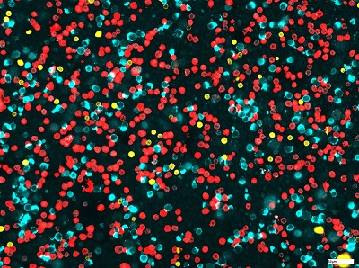
For chip cytometric analysis, the cells are applied onto a chip before they are stained using fluorescent dye-labeled antibodies that specifically bind to certain markers on the surface of the cells. This allows researchers to differentiate effectively between monocytes (light blue), T cells (yellow), and neutrophil granulocytes (red), for example. Courtesy of Fraunhofer ITEM.
For the analysis, patient samples are applied onto a special slide — in this case, a chip with a transparent chamber. The cells attach to the bottom of the chamber and then are stained using fluorescent dye-labeled antibodies that bind specifically to a certain type of immune cells. This allows researchers to dye, for example, granulocytes and macrophages different colors. Analysis of the microscopic images is highly automated, and the results allow the researchers to quantify the immune cells within the sample.
“Chip cytometry not only allows us to differentiate the cell types from each other, but also to see whether they are activated and what subgroup they belong to,” Carstensen said.
Chip cytometry was developed by Zellkraftwerk, a Hannover startup company. Zellkraftwerk was merged into Canopy Biosciences, which is actively developing commercial systems and other products for chip cytometry. Zellkraftwerk originally used blood cells to validate the technology. Carstensen adapted it for immune cells in the lungs.
In a pilot project in which Carstensen’s team showed how chip cytometry can be adapted to immune cells found in the lungs, the researchers triggered inflammation in healthy test subjects by having them inhale an irritant that mimics special components of the membrane surface of bacteria. Then they examined the subjects’ sputum and bronchoalveolar lavage fluid and used chip cytometry to track the lungs’ immune response.
“Cells that patients cough up in their sputum are often pre-activated, so that makes them harder to process than blood cells,” she said. The macrophages found in the lungs are also difficult to analyze, because they are much bigger than blood cells.
The team discovered elevated numbers of neutrophil granulocytes and monocytes in particular — that is, the cells that differentiate into macrophages in the lungs.
“We have demonstrated that we can detect inflammation and quantify it,” Carstensen said.
The project further highlighted advantages that chip cytometry offers when compared to traditional cell characterization methods. Unlike a differential cell count, whereby the different cell types are counted manually under a microscope, chip cytometry conducts the analysis automatically. The tool also has an edge on another standard method, flow cytometry, where samples are discarded after analyses.