Raman-based spectroscopy methods promise fast and label-free in vivo diagnosis.
IWAN W. SCHIE AND ALEKSANDAR LUKIC, LEIBNIZ INSTITUTE OF PHOTONIC TECHNOLOGY, AND JÜRGEN POPP, LEIBNIZ INSTITUTE OF PHOTONIC TECHNOLOGY AND INSTITUTE OF PHYSICAL CHEMISTRY, FRIEDRICH-SCHILLER UNIVERSITY
Spectroscopy has the potential to provide pathologists with fast in vivo tissue characterization to determine tumor type and grade and to delineate tumor margins. However, extending the applicability of microspectroscopy to in vivo imaging requires suitable optical fiber probes for an endoscopic inspection of difficult-to-access body regions. The engineering process is challenging. Besides the miniaturization of microscopy, it requires innovative scanning, excitation light delivery and signal collection, as well as robust and alignment-free light sources.
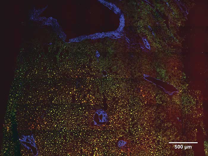
Figure 1. Multiphoton image of steatotic liver tissue using coherent anti-Stokes Raman scattering (CARS) microscopy acquired from the CH2 stretching vibration at 2850 cm-1. Visualizing lipid accumulations in the tissue, in red; the green signal is generated by two-photon excited autofluorescence (TPEF), highlighting retinol accumulations in the lipid droplets; the blue signal is from second-harmonic generation (SHG), highlighting the collagen matrix and the artery walls.
In recent years, spectroscopy-based methods, such as Raman spectroscopy, have seen a significant expansion from a standard tool in chemical analysis to state-of-the-art technology for medical diagnostics1,2. Besides spontaneous Raman spectroscopy, more sophisticated Raman-based modalities, such as coherent Raman scattering (CRS) microscopy or surface-enhanced Raman spectroscopy (SERS), have begun to find clinical application.
Raman spectroscopy is based on in-elastic light scattering between a photon and a molecule, resulting in the excitation of molecular vibrations. This excitation results in a characteristic Raman spectrum, providing a detailed molecular fingerprint about the molecular bonds present in the sample. Those can be correlated to more complex macromolecules such as proteins, lipids, nucleic acids, carbohydrates and others.
Raman spectroscopy frequently has been used for the label-free characterization of tumor tissue and single-cell analysis. Because of the ability to provide a precise molecular fingerprint of a sample, this family of methods is destined for clinical in vivo applications.
For in vivo applications, a fiber-based approach is necessary. In the simplest implementation, a fiber probe contains a central excitation core, surrounded by multiple collection fibers. The front of the fiber probe is covered by a composite filter that rejects the Raman signal generated in the excitation fiber, but allows the laser excitation to pass. The collection fibers are covered by a filter that allows the collection of the Raman signal, but rejects the excitation light. Such probes and modified versions of such probes have been widely used for grading of bladder and head and neck cancer, intraoperative brain tumor identification, arterial plaque compositions and other diagnostics3,4,5.
While spontaneous Raman spectroscopy provides a full molecular fingerprint of a sample, it is usually slow, and only local point information about the molecular composition can be measured. Hyperspectral Raman images are typically only acquired on ex vivo biopsies or single cells.
CRS microscopy methods, such as coherent anti-Stokes Raman scattering (CARS) microscopy and stimulated Raman scattering (SRS) microscopy, provide a significant improvement in terms of acquisition speed and throughput.
While the acquisition of a single Raman spectrum requires several seconds, a CARS or SRS signal can be acquired in microseconds and even nanoseconds, making it qualified to acquire large label-free images of tissue ex vivo and in vivo.
In most cases, a CRS signal is acquired from a single Raman band, or if from multiple bands, mostly bands from the high-wavenumber region. The implementation of CRS is frequently accompanied by signals based on other nonlinear optical processes such as two-photon excited autofluorescence (TPEF) and second-harmonic generation (SHG).
TPEF is generated from endogenous components of the extra-cellular matrix, such as elastin and NADP(H), but also from intracellular flavin coenzymes. The SHG signal, on the other hand, is generated in noncentrosymmetric molecular structures such as collagen or crystals. Today, there is rarely a study that only utilizes one of the methods.
Steatotic liver tissue, for example, has been imaged by multiphoton microscopy (Figure 1). The CARS signal, red in the figure, is acquired from the CH2 stretching vibration at 2850 cm−1, highlighting lipid accumulations in the tissue. The signal generated by TPEF (green in the figure), highlights retinol in the lipid droplets. The signal from SHG (blue in the figure) highlights the collagen matrix and the artery walls in the tissue.
While the technology for multiphoton microscopy platforms is fairly well established, the implementation of multiphoton modalities for in vivo diagnostics and in vivo tumor margin delineation using fiber probes remains challenging.
There are several aspects that have to be considered for development of an endoscopic fiber probe head. First, short-pulsed excitation light has to be delivered to the sample. This can be reasonably achieved for a single excitation beam, as is required for TPEF and for SHG.
The excitation of a CRS signal is not straightforward because of a difference in excitation wavelength that sometimes can be more than 200 nm. In addition, for CARS, a parasitic nonresonant background signal is generated in the fiber and can easily obscure the CARS signal from the sample. Filtering of the excitation light after the fiber is key. Great care has to be taken to maintain other optical properties of the excitation light, such as pulse width and phase relation between the excitation beams. These can change during the propagation in the fiber and affect the signal generation in the sample plane.
But even for TPEF and SHG, the short pulses experience a broadening, which affects the signal generation efficiency, and fluorescence signals can be generated within the fiber and obscure small signals.
The implementation of a scanning unit on a microscope is straightforward and is typically done by resonant or galvo-scanning mirrors. A highly sophisticated and widely used open source software for laser-scanning microscopes is ScanImage, which was developed at Janelia Farm and is now provided as open source through the Vidrio Technologies LLT (https://vidriotechnologies.com).
The implementation of a scanning unit in a fiber probe head, however, is orders of magnitude more complex. Because many in vivo applications require access to hard-to-reach regions, a significant miniaturization of the scanning unit is required. A great part of a microscopy platform has to be integrated into a probe head of a few millimeters in diameter, without loss of functionality. This is only possible with miniaturized scanning approaches such as microelectromechanical system (MEMS) scanners or piezo-tube scanners.
The typical edge length for MEMS scanner modules is on the order of a few millimeters with submillimeter size mirrors and can be found in both resonant and nonresonant forms. When both axes are based on resonant scanners, image reconstruction requires extra care. The light is scanned in a Lissajous pattern, and not all MEMS scanners provide a position feedback control.
Piezo-ceramic tubes for scanning are commercially available in different sizes and with inner diameters down to 0.8 mm, which significantly helps with the miniaturization of the probe head. Piezo tubes have segmented electrodes on opposite sides (four contacts) and an inner or outer ring electrode for ground.
When voltage is applied to the electrodes, the piezo tube contracts on one side and bends on the other, with a displacement of several µm. The delivery fiber is mounted in a collar inside the piezo tube, with a several-millimeter-long fiber piece extending out of the tube. When an oscillating voltage that matches the resonance frequency of the piezo tube with mounted fiber is applied, the fiber can exhibit a rapid displacement. When the voltage is alternatingly applied to the orthogonal directions of the tube, a spiral pattern can be produced and used to perform a fiber scanning of the sample.
Both MEMS-based and piezo-based implementations require significant engineering know-how for a good performance. An approach that was recently introduced6 circumvents the complications with the miniaturization of the scanning unit by relying for excitation light delivery on imaging fibers, which are commonly used in endoscopes. The scan is performed at the distal end of the imaging fiber, using conventional galvo-scanning mirrors.
The fiber probe head was developed by Grintech GmbH of Jena, Germany. The implementation significantly reduces the complications of fabricating a small scanner unit inside a probe head and enables multiphoton imaging of, for example, skin tissue, using CARS, TPEF and SHG (Figure 2).
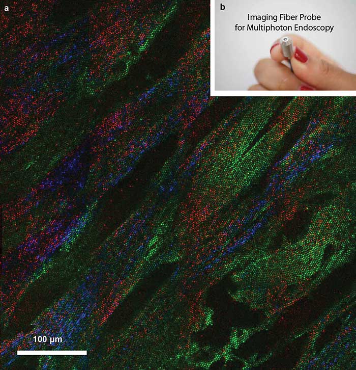
Figure 2. Multimodal image acquired by a multiphoton imaging-fiber probe of human skin composed of keratin and lipids in the upper primary layer (epidermis), and collagen and elastin fibers in the extracellular matrix (a). The TPEF signal of keratin is shown in green, the CARS signals of epidermis in red, and the SHG signal of collagen fibers in blue. The compact multiphoton imaging fiber-based endoscope used in this application (b).
Common multiphoton microscopy setups, which include a laser-scanning microscope and a laser unit, are large and can easily take up an entire optical bench, making the transition to a clinical environment difficult. Besides the compact implementation of a scanning unit, the miniaturization of the excitation source is crucial.
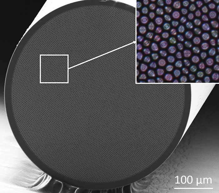
Figure 3. Imaging fiber used for the multiphoton microscopy implementation by Lukic et al. See reference 6.
For the generation of a CRS signal, two synchronized short high-power pulses are required. One of the excitation beams is used as the Stokes beam and to pump an optical parametric oscillator (OPO), which is then used as the pump beam to generate the CRS process. The use of an OPO allows continuous scanning of the pump wavelength and the probe of an extended spectra region. Fiber-based lasers represent a significant development for CRS applications. The lasers are highly compact and provide at the output a readily overlapped pump-probe beam combination. This allows reduction of the optical setup for beam combination and shaping to a minimum and ensures further miniaturization of the system7.
Meet the authors
Iwan W. Schie received a Ph.D. in biomedical engineering from the University of California, Davis. His research focus was on instrumental development of multiphoton microscopy and spontaneous Raman spectroscopy for medical applications. From 2013 to mid-2017, he was a postdoctoral researcher at the Leibniz Institute of Photonic Technology in Jena, Germany, developing high-throughput Raman spectroscopy systems for single-cell classification. He is now leading a research group for multimodal instrumentation at the Leibniz-IPHT; email: [email protected].
Aleksandar Lukic studied microsystems engineering at the University of Applied Sciences in Regensburg, Germany, and laser and optotechnologies at the Ernst Abbe University of applied sciences in Jena. In 2013, he joined the group of professor Jürgen Popp at the Institute of Physical Chemistry at the Friedrich Schiller University Jena, where he focuses on fiber probes for nonlinear imaging applications; email: [email protected].
Jürgen Popp holds a chair for physical chemistry at the Friedrich-Schiller University Jena. He is also the scientific director of the Leibniz Institute of Photonic Technology, Jena. His research interests are mainly concerned with biophotonics. In particular, his expertise is in the development and application of innovative Raman techniques for biomedical diagnosis; email: [email protected].
Acknowledgments
We gratefully acknowledge financial support from the EU, the “Thüringer Ministerium für Wirtschaft, Wissenschaft und Digitale Gesellschaft,” the “Thüringer Aufbaubank,” the Federal Ministry of Education and Research, Germany (BMBF), the German Science Foundation, the Fonds der Chemischen Industrie and the Carl-Zeiss Foundation are gratefully acknowledged.
References
1. C. Krafft et al. (2015). Developments in spontaneous and coherent Raman scattering microscopic imaging for biomedical applications. Chem Soc Rev, Vol. 45, pp. 1819-1849.
2. C. Krafft et al. (2017). Label-free molecular imaging of biological cells and tissues by linear and non-linear Raman spectroscopic approaches. Angew Chem Int Ed, Vol. 56, pp. 4392-4431.
3. C. Krafft et al. (2012). Diagnosis and screening of cancer tissues by fiber-optic probe Raman spectroscopy. Biomed Spectrosc Imaging, Vol. 1, Issue 1, pp. 39–55.
4. I. Latka et al. (2013). Fiber optic probes for linear and nonlinear Raman applications — Current trends and future development. Laser Photon Rev, Vol. 7, Issue 5, pp. 698–731.
5. M. Jermyn et al. (2015). Intraoperative brain cancer detection with Raman spectroscopy in humans. F Sci Transl Med, Vol. 7, Issue 274, pp. 274ra19.
6. A. Lukic et al. (2017). Endoscopic fiber probe for nonlinear spectroscopic imaging. J Optica, Vol. 4, Issue 5, p. 496.
7. T. Gottschall et al. (2015). Fiber-based light sources for biomedical applications of coherent anti-Stokes Raman scattering microscopy. Laser Photon Rev, Vol. 9, Issue 5, pp. 435–451.